The Nobel Prize in Chemistry 2017 was awarded to Jacques Dubochet, Joachim Frank, and Richard Henderson for their development of Cryo-Electron Microscopy (cryo-EM), which simplified and improved the imaging of biomolecules’ structure.
Did you notice, when you look at biochemical related news a certain period of time before, the pictures of the virus or other biomolecular structure just looked like the rough photos in old 3D horror games, or we say fake 3D models. But it seems to be quite different recent years? Yes, the year of 2013 is the turning point. Scientists can start with a microscope to see high-resolution biomolecule structure, and even with rotate 3-D, and this is supported by the revolution of Cryo-Electron Microscopy.
Since the invention of microscope, scientists are all expecting to the improvement of technologies to see the more subtle molecular structures. Optical microscope enables the observation of cell structures, for example, to see the location of proteins in the cells; and electron microscopy enables more detailed imaging, such as the molecular structure of proteins. But the problem is: the electron beam will cause radiation damage to the biological material. This may recall the words in high school physics that scientific observation should try to avoid disturbing subjects so as not to lead to inaccurate or even incorrect results. That’s why electron microscopes were long believed only to be suitable for imaging dead matters instead of biological materials.
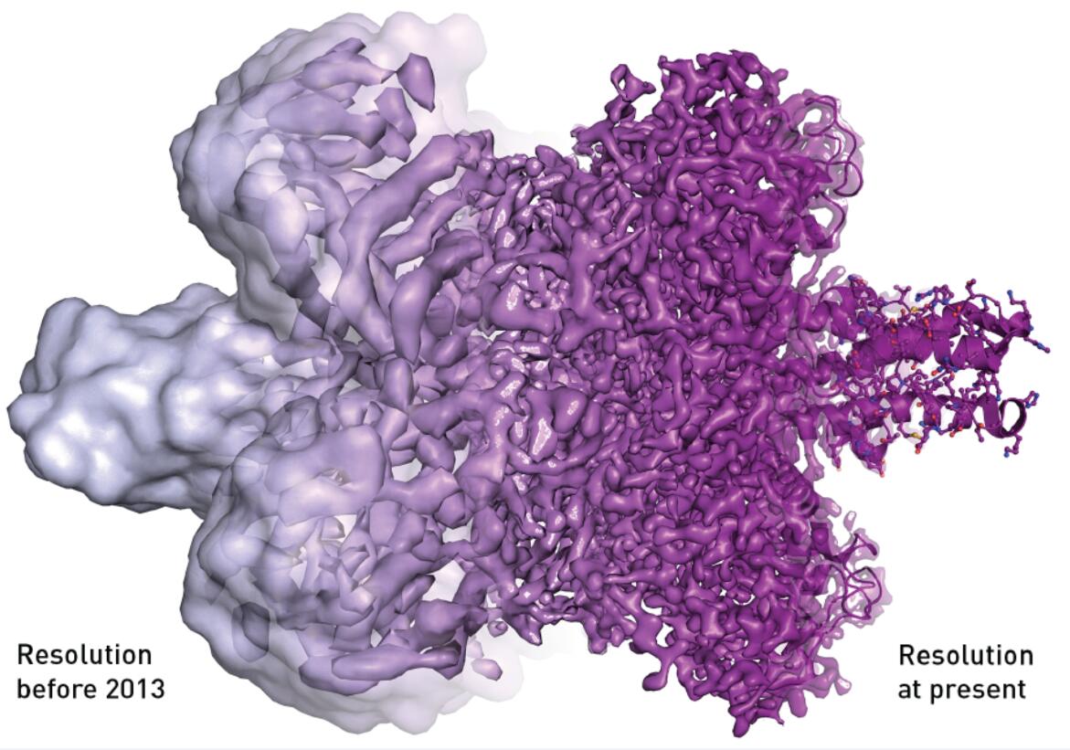
Actually, the initial development of Electron microscopy can be dated back to 1931. And until the late 1980s, Cryo-Electron Microscopy was developed. However, at that time, X-ray was the popular star and the Cryo-Electron Microscopy was still in its embryonic stage. No one was optimistic about the technology except the pioneer Henderson. He predicted that “Cryo-Electron Microscopy will go beyond other technologies and become the main force for the study of protein structure.” And at the year of 1990, Richard Henderson succeeded in using an electron microscope to generate a 3D image of a protein at atomic resolution.
The contribution of Frank can be attributed to the development of image processing method. Different from the observation of stones, metal and other static objects, observation of biological samples is quite difficult. Because the traditional method only obtain plane image and are quite vague. Frank combines multiple fuzzy two dimensional shadows from an electron microscope diffraction pattern into a single three dimensional structure.
What’s more interesting is the contribution of Jacques Dubochet. To avoid the biological material being burned, people used to utilize icing method. But what’s came along was the problem that the frozen ice crystal structure would make the image distorted. And the observation of the movement of molecules would be hindered in the ice state. To solve this problem, Dubochet came up with a new idea. He puts the sample in to liquid ethane and helps the molecules keep their shape and movement. No burn, no ice crystal. Then we can get a clear image without distortion.
Thanks to those amazing discoveries, observations of many virus come to the atomic level, bringing new hope for the research of some diseases considered incurable. Biochemistry is now facing an explosive development and is all set for an exciting future.