
Uncategorized Wednesday, 2026/10/21
Research Highlights
A landmark study published in Nature provides a comprehensive view of how the human prefrontal cortex changes throughout life. By analyzing more than 1.3 million cell nuclei from neurologically normal individuals, the PsychAD Consortium constructed a lifespan-wide single-nucleus transcriptomic atlas of the human prefrontal cortex. The findings reveal three major phases of molecular change—early development, relative stability during midlife, and selective reactivation of molecular programs in late life—offering new insights into brain aging, cellular resilience, and age-related neurological disease vulnerability.
Mapping the Human Prefrontal Cortex Across the Lifespan
The dorsolateral prefrontal cortex (DLPFC) is a critical brain region responsible for higher-order cognitive functions, including executive control, working memory, decision-making, and behavioral regulation.
It is also among the brain regions particularly vulnerable to age-related changes in cognition and neurological function.
Previous investigations into human brain development and aging have often focused on specific periods, such as childhood, adulthood, or advanced age, or on brain tissues affected by neurological disease.
Although these studies have provided valuable insights, they have offered only fragmented snapshots of the molecular processes occurring throughout the human lifespan.
As a result, researchers have lacked a unified framework for understanding how cellular and transcriptional programs evolve continuously from early brain development through adulthood and into old age.
A comprehensive lifespan atlas is therefore essential for identifying the molecular transitions that distinguish normal maturation, stable adult function, and age-associated vulnerability.
Nature Study Analyzes More Than 1.3 Million Cell Nuclei
A research team led by Panos Roussos at the Icahn School of Medicine at Mount Sinai, with Hui Yang as the first author, published a study in Nature entitled “Lifespan single-cell transcriptomic atlas of the human prefrontal cortex.”
Working through the PsychAD Consortium, the researchers analyzed more than 1.3 million cell nuclei obtained from neurologically normal individuals spanning the human lifespan.
Using single-nucleus RNA sequencing (snRNA-seq), they developed a comprehensive transcriptomic reference of the human prefrontal cortex.
This approach enabled the researchers to characterize gene expression patterns across major brain cell populations and investigate how individual cell types change with age.
Rather than examining brain aging as a single, uniform process, the study resolved age-related molecular trajectories at the cellular level, revealing distinct patterns associated with neurons, glial cells, and other brain cell populations.
The resulting atlas provides a systematic reference for investigating the molecular organization of the human brain across different stages of life.
Three Major Phases of Molecular Change During Brain Aging
One of the most important findings was that molecular changes in the prefrontal cortex do not follow a simple linear trajectory.
Instead, the researchers identified three broad phases of transcriptional change across the human lifespan.
Early Life: Extensive Neuronal Reprogramming and Circuit Development
During early development, neurons undergo substantial transcriptional remodeling as the brain establishes and refines its functional networks.
Gene expression programs associated with neuronal maturation, synaptic organization, and cellular adaptation are actively regulated during this period.
These molecular changes support the formation and refinement of neuronal connections required for increasingly complex cognitive functions.
The early developmental stage is therefore characterized by considerable molecular plasticity and dynamic cellular adaptation.
Midlife: A Period of Relative Transcriptomic Stability
Following the extensive changes of early development, the prefrontal cortex enters a comparatively stable molecular phase during adulthood and midlife.
Across many cell populations, gene expression patterns show relatively limited changes compared with the pronounced transcriptional remodeling observed during development.
This period resembles a molecular plateau, during which established cellular programs are largely maintained.
Importantly, relative transcriptional stability does not imply the complete absence of biological aging. Rather, it indicates that major cell-type-specific gene expression programs remain comparatively consistent during this stage.
Late Life: Selective Reactivation of Molecular Programs
In advanced age, certain molecular pathways undergo renewed transcriptional changes.
Rather than exhibiting uniform deterioration across all cellular functions, the aging prefrontal cortex displays selective reactivation and reorganization of specific biological programs.
These changes differ across cell types and may involve stress responses, immune-related signaling, and cellular regulatory networks.
The findings suggest that brain aging is not a constant, evenly progressing process. Instead, it includes periods of relative stability followed by more pronounced molecular transitions.
This nonlinear pattern may help researchers identify stages of life during which the brain becomes increasingly susceptible to age-associated dysfunction.
Neurons and Glial Cells Follow Distinct Aging Trajectories
The study also demonstrated that different brain cell types exhibit distinct molecular responses to aging.
In neurons, stress-resilience-related transcriptional programs become particularly active during early life, potentially helping cells adapt to the substantial physiological demands associated with brain development.
These programs subsequently become more stable as the brain reaches maturity.
Glial cells, by contrast, show more pronounced molecular alterations during late life.
These changes include increased immune-related activity, enhanced cellular stress responses, and reorganization of genes associated with circadian regulation.
This distinction highlights the importance of studying brain aging at the level of individual cell populations.
Neurons and glial cells do not necessarily experience the same molecular transitions at the same stages of life, even though they interact closely to maintain brain function.
Furthermore, these age-associated programs are not randomly distributed throughout the brain.
The researchers identified anatomically organized expression patterns across cortical layers and between gray and white matter, indicating that molecular aging is influenced by spatial and cellular context.
Circadian Gene Expression Undergoes Reorganization in the Aging Brain
Another notable discovery involved changes in circadian-related gene expression during advanced aging.
Circadian rhythms regulate numerous physiological processes, including sleep–wake cycles, metabolism, hormone secretion, and cellular stress responses.
The study identified widespread reorganization of genes associated with 24-hour biological rhythms in the aging prefrontal cortex.
In neuronal populations, the coordinated expression of circadian-related genes became less synchronized, suggesting a decline in the coherence of internal molecular timing programs.
Meanwhile, glial cells exhibited altered rhythmic patterns associated with cellular stress responses.
These observations reveal that age-related disruption of biological timing may involve distinct molecular mechanisms in neurons and glial cells.
The findings are particularly relevant because sleep disturbances and fluctuations in cognitive performance are common among older adults.
Although the study does not establish that the observed transcriptional changes directly cause these clinical symptoms, it suggests that circadian dysregulation may be an important component of the transition from cellular resilience toward increased vulnerability during aging.
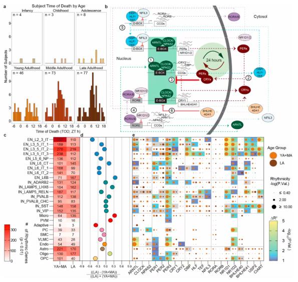
Fig 1. Age-associated changes in 24-hour gene expression rhythms in the human prefrontal cortex.
Linking Normal Brain Aging to Neurological and Psychiatric Disease Risk
To investigate the potential relevance of the atlas to human disease, the researchers integrated age-associated gene expression modules with genetic risk factors implicated in psychiatric and neurodegenerative disorders.
This analysis revealed that certain disease-associated genes overlap with molecular programs that become reactivated or reorganized during late life.
Such relationships may help explain why genetic susceptibility to some neurological conditions does not necessarily result in disease during early adulthood but becomes more clinically relevant with advancing age.
For example, a genetic variant may remain relatively well tolerated during periods of stable cellular function but contribute to vulnerability when age-associated stress responses or regulatory networks undergo substantial changes.
These findings provide a molecular framework for examining interactions between inherited disease risk and the biological processes of aging.
They also suggest that disease susceptibility should be understood in the context of both cell type and developmental stage.
Importantly, the atlas was constructed using neurologically normal individuals, making it a valuable reference for distinguishing physiological aging processes from disease-associated molecular alterations in future investigations.
A Reference Framework for Future Brain Aging Research
The significance of this study extends beyond the identification of age-related gene expression changes.
By establishing a large-scale, cell-type-resolved transcriptomic atlas spanning the human lifespan, the PsychAD Consortium has created a reference framework for studying normal brain development, maturation, and aging.
This resource may support future investigations into several important questions: when age-associated molecular vulnerability begins to emerge, which brain cell populations are particularly susceptible to specific diseases, and how genetic risk factors interact with cellular aging programs.
The atlas may also help researchers identify potential windows for intervention before age-associated molecular changes become associated with substantial neurological dysfunction.
However, these possibilities require further functional validation and longitudinal investigation.
Because the study primarily analyzes molecular profiles across individuals of different ages, the inferred lifespan trajectories should not be interpreted as direct longitudinal measurements of the same individuals over time.
Nevertheless, its scale and cellular resolution represent an important advance in understanding the molecular architecture of human brain aging.
Conclusion: Brain Aging Is a Dynamic, Nonlinear Process
This study reveals that molecular aging in the human prefrontal cortex follows a complex, nonlinear trajectory rather than a uniform process of progressive decline.
By analyzing more than 1.3 million cell nuclei, the researchers identified three major phases of transcriptional change: extensive remodeling during early development, relative stability throughout midlife, and selective molecular reorganization in advanced age.
The findings also demonstrate that neurons and glial cells follow distinct aging trajectories, with important differences in stress-response regulation, immune-related signaling, and circadian gene expression.
Furthermore, the overlap between age-associated molecular programs and genetic risk factors for neurological disorders offers new insights into how normal brain aging may influence disease susceptibility.
Overall, the lifespan transcriptomic atlas provides a valuable cellular and molecular reference for investigating human brain development, aging, and age-related neurological disease.
Related Products & Services
- Cell and Gene Therapy
- Immune Checkpoint Proteins
- ADC Target Protein
- PROTAC Targets
- Targets of CAR-T Cell Therapy
- Cancer Drug Targets
- Protein Engineering Services
- Protein Interaction Service
- Protein Expression and Purification Services
- Drug Discovery Screening
- Protein Pathway Profiling
Reference
Yang, H., Clarence, T., Scott, M.R. et al. Lifespan single-cell transcriptomic atlas of the human prefrontal cortex. Nature 657, 1003–1015 (2026). https://doi.org/10.1038/s41586-026-10271-7