
Uncategorized Wednesday, 2026/10/21
Research Highlights
A groundbreaking study published in Nature provides a comprehensive spatial reference framework for understanding human heart development and the molecular mechanisms underlying congenital heart disease (CHD). By integrating multiple single-cell and spatial omics technologies, researchers constructed a detailed atlas of the developing human heart and identified cellular abnormalities associated with trisomy 21 (T21), also known as Down syndrome. The findings offer new insights into how disrupted cellular organization and developmental signaling may contribute to congenital cardiac abnormalities.
Understanding Human Heart Development and Congenital Heart Disease
Human heart development is a highly coordinated biological process that requires precise spatial and temporal interactions between cells derived from the cardiac mesoderm and neural crest.
Together, these cell populations contribute to the formation of essential cardiac structures, including the sinoatrial node, and support the progressive maturation of the myocardium.
Disruptions in these developmental processes can lead to congenital heart disease (CHD), one of the most common categories of congenital abnormalities.
Individuals with trisomy 21 (T21), commonly known as Down syndrome, have a significantly elevated risk of developing congenital heart defects. This increased susceptibility highlights the sensitivity of cardiac development to gene dosage and chromosomal imbalance.
However, researchers have long lacked a sufficiently comprehensive spatial reference map to explain how early molecular disturbances translate into abnormalities in tissue organization and cardiac structure.
Understanding these spatial relationships is essential for identifying the cellular and molecular mechanisms that connect genetic alterations to congenital heart defects.
Nature Study Establishes a Multimodal Atlas of Human Cardiogenesis
A research team led by Sarah A. Teichmann at the University of Cambridge published a study in Nature entitled “Dynamics of human cardiogenesis and its disruption in trisomy 21.”
The study addresses a major gap in developmental biology by constructing a multimodal spatial atlas of the human heart across developmental stages ranging from 4 to 20 weeks post-fertilization.
To achieve this, the researchers integrated several complementary technologies, including:
- Single-cell transcriptomics: To characterize gene expression programs across distinct cardiac cell populations.
- Single-nucleus DNA methylation profiling: To investigate epigenetic regulatory patterns during cardiac development.
- Single-nucleus ATAC sequencing (snATAC-seq): To assess chromatin accessibility and identify regulatory regions associated with cell differentiation and maturation.
- Spatial transcriptomics: To map gene expression patterns to their anatomical locations within developing cardiac tissues.
By integrating these datasets, the researchers identified 21 distinct tissue microenvironments within the developing human heart.
They also developed a hierarchical classification tool called TissueTypist, which enables automated annotation of tissue microenvironments at different spatial resolutions.
The accuracy and robustness of TissueTypist were evaluated using histological analyses and independent datasets.
This integrated approach provides a systematic framework for examining how cardiac cell populations emerge, interact, and organize into functional structures throughout human development.
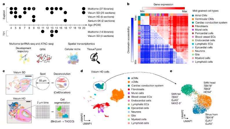
Fig 1. Multimodal spatial atlas of the developing human heart.
Spatial Organization of Pacemaker Cells and Cardiac Innervation
The newly established atlas revealed previously underappreciated complexity in the development of the cardiac conduction system.
One particularly important finding concerned the sinoatrial node, the heart's primary pacemaker responsible for initiating rhythmic electrical impulses.
Rather than forming a homogeneous population, pacemaker cells within the sinoatrial region were organized into three distinct subtypes distributed along a linear spatial axis.
During early gestation, pacemaker cell subtypes located in the sinus venosus region and the head of the sinoatrial node exhibited molecular programs associated with neural attraction.
These cells interacted with parasympathetic neurons through signaling pathways involving Eph–ephrin and semaphorin–plexin interactions.
Such molecular communication suggests that the spatial coordination between the developing cardiac pacemaker system and autonomic neural inputs is established at an early developmental stage.
The findings provide important insights into how electrical conduction structures and neural regulatory networks become integrated during human cardiogenesis.
Our Featured Proteins


Molecular Gradients Regulate Ventricular Myocardial Maturation
The atlas also identified distinct cellular and transcriptional gradients across the ventricular wall.
These gradients were distributed along both the inner-to-outer anatomical axis and a computationally inferred developmental pseudotime trajectory.
The observed spatial and temporal patterns provide a molecular framework for understanding ventricular myocardial compaction, a critical developmental process through which the ventricular myocardium acquires its mature structural organization.
During normal cardiac development, the ventricular wall undergoes extensive remodeling as cardiomyocytes proliferate, differentiate, and progressively organize into compact myocardial layers.
Disruption of these coordinated processes can compromise ventricular architecture and cardiac function.
By linking transcriptional states to specific spatial locations, the study offers new insights into the molecular programs that support myocardial maturation and ventricular wall development.
Trisomy 21 Disrupts Cardiomyocyte Survival and Myocardial Compaction
A particularly significant aspect of the study involved comparing developing hearts from individuals with trisomy 21 with age-matched control samples.
The researchers identified pronounced abnormalities in ventricular cardiomyocyte populations in T21 hearts.
Specifically, the number of compact ventricular cardiomyocytes (vCMs) was reduced, while markers of apoptosis were significantly elevated.
These findings suggest that impaired cardiomyocyte survival may contribute to defective myocardial development in trisomy 21.
To further investigate this relationship, the researchers used induced pluripotent stem cell (iPSC)-derived cardiomyocytes generated from genetically matched cellular models.
The T21 cardiomyocytes exhibited increased apoptosis, reproducing a key pathological feature identified in the developing heart tissues.
These observations provide additional evidence that trisomy 21 can directly influence cardiomyocyte survival and developmental programs.
Importantly, the findings suggest that the increased risk of CHD associated with Down syndrome may arise from cumulative disturbances in early cardiomyocyte survival and myocardial compaction rather than from the effects of a single gene mutation.
The study therefore highlights the importance of chromosome dosage imbalance in shaping cardiac developmental trajectories.
A Reusable Framework for Investigating Congenital Heart Disease
Beyond generating a detailed anatomical and molecular atlas, the researchers established a reusable analytical framework for studying human developmental disorders.
The integration of spatial transcriptomics, single-cell profiling, and epigenomic information makes it possible to investigate disease-associated molecular changes within defined tissue microenvironments.
In future studies, this approach could be combined with patient-specific genetic backgrounds, environmental exposures, and experimental drug interventions to explore the developmental origins of congenital heart defects.
Rather than examining individual genes or cell types in isolation, researchers may be able to trace how molecular disturbances emerge and spread across developing tissues.
The TissueTypist classification framework also has potential applications beyond cardiac development.
With appropriate adaptation and validation, similar analytical strategies could be applied to other developing organs to investigate tissue organization, cell differentiation, and disease-associated developmental abnormalities.
Future Research Directions and Clinical Relevance
Although the study represents a major advance in understanding human cardiogenesis, several questions remain unresolved.
The current atlas primarily focuses on normal cardiac development and developmental alterations associated with trisomy 21.
Additional investigations involving other congenital heart disease subtypes will be necessary to determine whether similar cellular and molecular mechanisms contribute to a broader range of cardiac abnormalities.
Furthermore, more extensive developmental time-course studies and functional validation experiments are needed to clarify the causal relationships between specific molecular changes and structural cardiac defects.
Nevertheless, the findings provide an important foundation for future investigations into congenital heart disease.
By establishing a systematic spatial reference framework for human heart development, the study enables researchers to examine how genetic alterations affect cardiac cell populations, tissue organization, and developmental signaling networks.
Conclusion
This research represents an important step toward understanding the cellular and molecular dynamics of human heart formation.
The comprehensive multimodal atlas reveals previously unrecognized spatial organization of pacemaker cells, developmental gradients associated with ventricular myocardial maturation, and abnormalities in cardiomyocyte survival linked to trisomy 21.
By combining high-resolution spatial information with single-cell and epigenomic profiling, the study establishes a valuable reference for investigating the developmental origins of congenital heart disease.
More broadly, it demonstrates the potential of integrated spatial omics technologies to advance our understanding of human organ development and congenital disorders.
Related Products & Services
- Cell and Gene Therapy
- Immune Checkpoint Proteins
- ADC Target Protein
- PROTAC Targets
- Targets of CAR-T Cell Therapy
- Cancer Drug Targets
- Protein Engineering Services
- Protein Interaction Service
- Protein Expression and Purification Services
- Drug Discovery Screening
- Protein Pathway Profiling
Reference
Cranley, J., Kanemaru, K., Bayraktar, S. et al. Dynamics of human cardiogenesis and its disruption in trisomy 21. Nature (2026). https://doi.org/10.1038/s41586-026-11125-y