
Uncategorized Thursday, 2026/08/06
This study establishes a DG–RSGc–NPY–IDO1–L-Kyn signaling axis, elucidating the molecular mechanism by which central neural circuit activity regulates fracture repair. It further identifies the peripheral components of this pathway as potential therapeutic targets for impaired bone healing.
Traumatic brain injury is commonly associated with accelerated fracture repair; however, the neural circuits linking brain status to skeletal healing have remained unclear.
Li Weixu from Zhejiang University and Wang Yongjie from Hangzhou Normal University published a research article entitled “A circuit linking dentate gyrus and retrosplenial granular cortex regulates fracture healing in male mice” online in Nature Communications.
The study demonstrates that, in male mice, a hippocampal–cortical circuit connecting the dentate gyrus (DG) and the retrosplenial granular cortex (RSGc) regulates the repair process following bone injury.
Bone injury simultaneously increased neuronal activity-associated signals in both brain regions. Inhibition of excitatory neurons in either the DG or RSGc, or disruption of neuronal populations associated with the DG–RSGc pathway, accelerated bone repair. Conversely, activation of excitatory neurons in the DG or RSGc delayed healing.
Activation of the RSGc reversed the pro-healing effect caused by DG inhibition, indicating that, under the experimental conditions used, the regulation of bone repair depended on the DG–RSGc neural circuit.
Following inhibition of this circuit, circulating levels of neuropeptide Y (NPY) increased, expression of indoleamine 2,3-dioxygenase 1 (IDO1) was upregulated in macrophages at the injury site, and the concentration of L-kynurenine (L-Kyn) was elevated. This metabolite promoted osteogenic differentiation.
Pharmacological blockade and rescue experiments confirmed the existence of an NPY–IDO1–kynurenine effector axis that links central neural circuit activity to peripheral skeletal repair.
Our Featured Proteins
| Cat.No. # | Product Name | Source (Host) | Species | Tag | Protein Length | Price |
|---|---|---|---|---|---|---|
| NPY-1350H | Recombinant Human NPY, GST-tagged | E.coli | Human | GST | 1-97aa | |
| NPY-233H | Recombinant Human NPY protein, His-tagged | E.coli | Human | His |

|
|
| NPY-262H | Recombinant Human NPY protein, Fc-tagged | HEK293 | Human | Fc | 29-64 aa |
|
| IDO1-14054H | Recombinant Human IDO1, GST-tagged | E.coli | Human | GST | 1-403a.a. | |
| Ido1-2126M | Recombinant Mouse Indoleamine 2,3-dioxygenase 1, His-tagged | E.coli | Mouse | His | 1-407 a.a. | |
| IDO1-3920H | Active Recombinant Human IDO1 protein, His-tagged | E.coli | Human | His | 2-403 aa | |
| IDO1-7190H | Recombinant Human IDO1 protein | E.coli | Human | Non | 2-403 aa | |
| IDO1-7325H | Recombinant Human IDO1, None tagged | E.coli | Human | Non | 1-403 a.a. |
Fracture Healing and Brain–Bone Communication
Fractures are common traumatic injuries, and approximately 5% to 10% of all fracture cases develop delayed union or nonunion. As private vehicle ownership increases and populations continue to age, the annual incidence of fractures is rising, while the body’s intrinsic healing capacity declines with age.
Delayed fracture healing and nonunion impose substantial physical and psychological burdens on patients. Repeated surgical procedures and long-term care also create considerable economic and social costs.
Therefore, clarifying the mechanisms underlying fracture healing and developing interventions that reduce the incidence of delayed union and nonunion have clear clinical value.
Traditionally, fracture healing has been regarded as a purely local repair process. However, growing clinical evidence indicates that patients with fractures accompanied by traumatic brain injury (TBI) often exhibit faster fracture healing and greater callus formation than patients with isolated fractures.
This phenomenon suggests the existence of important brain–bone interactions, although the underlying neural mechanisms remain poorly understood.
TBI can reshape systemic immune status and autonomic neural output, thereby regulating bone marrow activity and macrophage phenotype at peripheral injury sites.
In addition to autonomic regulation, multiple experimental models have implicated several other mechanisms in brain-to-bone communication, including:
- humoral and endocrine mediators;
- autonomic and sensory neural pathways;
- neuroimmune interactions;
- extracellular vesicles and small extracellular vesicles carrying osteogenic signals.
Bone metabolic homeostasis is a dynamic physiological process regulated by central signals from the brain. Large-scale genetic studies have demonstrated associations between human brain-related traits and bone mineral density.
Hypothalamic neurons integrate peripheral signals and convert them into physiological outputs that regulate metabolism and skeletal homeostasis.
Multiple hypothalamic pathways control skeletal homeostasis through autonomic and endocrine outputs, whereas stress-associated neural circuits can induce bone loss through the peripheral sympathetic nervous system.
Together, these findings demonstrate a complex relationship between central nervous system function and skeletal health. However, unlike the mechanisms regulating basal bone metabolism, the specific neural circuits that directly connect defined brain regions to fracture repair, as well as their downstream immunometabolic mediators, have not been fully characterized.
DG–RSGc Circuit Activity Controls Bone Repair
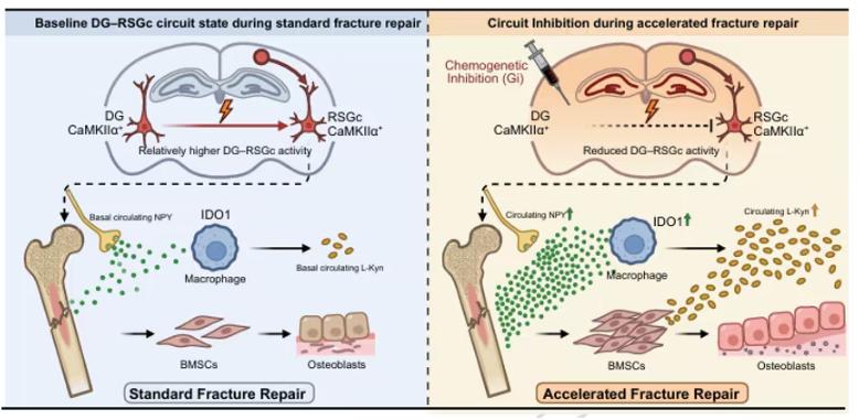
Graphical Abstract (Nature Communications)
Using multiple complementary bone injury models, together with neuronal activity labeling, viral circuit tracing, and bidirectional chemogenetic manipulation of targeted projections, the researchers identified a hippocampal–cortical circuit connecting the dentate gyrus and the retrosplenial granular cortex.
The study demonstrated that activity within the DG–RSGc circuit regulates the progression of skeletal repair in a proximal tibial defect (PTD) model.
Untargeted serum metabolomics and targeted metabolic profiling revealed extensive remodeling of tryptophan metabolism. Increased L-kynurenine emerged as a characteristic downstream feature of this process.
Subsequent experiments identified a neuroimmune–metabolic cascade in which suppression of neural circuit activity increased circulating NPY levels. Elevated NPY induced IDO1 expression in callus-associated macrophages, resulting in local accumulation of L-Kyn and subsequent promotion of osteogenic differentiation.
The study also employed an impact-induced TBI model, in which skeletal repair was markedly accelerated. Circulating NPY and L-Kyn levels increased simultaneously, demonstrating that these peripheral effector signatures were not limited to local circuit manipulation and may represent a more broadly applicable response.
Overall, this study establishes a DG–RSGc–NPY–IDO1–L-Kyn signaling axis, providing a mechanistic explanation for how central neural circuit activity controls fracture repair. It further suggests that the peripheral components of this pathway may serve as potential intervention targets for delayed bone healing and other bone repair disorders.
Related Products & Services
- Cell and Gene Therapy
- Immune Checkpoint Proteins
- ADC Target Protein
- PROTAC Targets
- Targets of CAR-T Cell Therapy
- Cancer Drug Targets
- Protein Engineering Services
- Protein Interaction Service
- Protein Expression and Purification Services
- Drug Discovery Screening
- Protein Pathway Profiling
Reference
- Zhang, W., Zhang, W., Guo, Y., Chen, D., Jiang, X., Zhang, J., Chen, E., Yuan, Y., Wu, W., Qian, Y., Yu, X., Wang, Y., & Li, W. (2026). A circuit linking dentate gyrus and retrosplenial granular cortex regulates fracture healing in male mice. Nature Communications. https://doi.org/10.1038/s41467-026-75340-x