
Uncategorized Monday, 2026/09/14
6. Mitotic Chromosome Condensation Is a Reversible Volume Phase Transition
DOI: 10.1126/science.adz1083
When cells enter mitosis, chromosomes undergo two major transformations: they become organized into cylindrical chromatids and condense to densities approaching those of protein crystals.
Loop extrusion driven by the condensin family may explain the formation of the cylindrical chromosome architecture. The mechanism underlying chromosome condensation itself, however, has remained unresolved.
When condensin is removed, chromosomes collapse into amorphous masses whose density is indistinguishable from that of normal mitotic chromosomes.
This observation suggests that cylindrical organization and condensation are two distinct processes.
A new study investigated the physical mechanism underlying chromosome condensation.
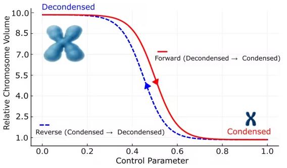
Researchers developed a system that allowed purified mitotic chromosomes to undergo reversible decondensation and recondensation in a parallel-plate flow chamber under optical microscopy.
This setup enabled precise control of the chromosome environment while chromosome morphology was directly observed.
Automated chromosome morphometry allowed high-throughput quantitative analysis of thousands of individual chromosomes.
The researchers applied classical polyelectrolyte-gel theory to chromatin, incorporating mixing energy, elastic energy, and Donnan free-energy terms, together with an explicit counterion-binding term adapted for chromatin.
By fitting the experimental dataset to the gel model, they tested whether the theory could account for chromosome condensation behavior.
Chromosome swelling and contraction were completely isotropic in three dimensions and reproducible over multiple cycles, with unchanged endpoint morphologies.
The chromatin-adapted classical polyelectrolyte-gel theory accurately predicted condensation curves under a wide range of conditions.
The theory also produced a surprising prediction: chromosomes swollen by lowering ionic strength should recondense at extremely low ionic strength.
This prediction was experimentally confirmed.
At high ionic strength, the chromosomes swelled again, displaying so-called reentrant behavior.
The experiments also revealed hallmarks of a two-state system, including phase coexistence within chromosomes and hysteresis during decondensation-recondensation cycles.
The gel model predicted that the two states interconvert through a volume phase transition, or VPT.
Under physiological ionic strength, mitotic chromosomes appeared to lie very close to this phase-transition threshold.
7. A Molecular Switch in TRAK Proteins Controls Directional Mitochondrial Transport in Response to Cellular Stress
DOI: 10.1126/science.aeh1475
Eukaryotic cells rely on microtubules as intracellular transport tracks for long-distance movement of cellular cargo.
Two major classes of molecular motors—kinesin and dynein—generally move in opposite directions along microtubules. Kinesin transports cargo toward the microtubule plus end, usually toward the cell periphery, whereas dynein transports cargo toward the minus end, generally toward the cell center.
Mitochondria, the major energy-producing organelles of the cell, must be dynamically distributed according to local energy demands.
Healthy mitochondria are transported toward regions of high energy consumption, such as neuronal synapses or sites of muscle contraction, whereas damaged mitochondria may be transported back toward the cell center for autophagic recycling.
How cells coordinate the activities of opposing kinesin and dynein motors on the same mitochondrial cargo has long remained unresolved.
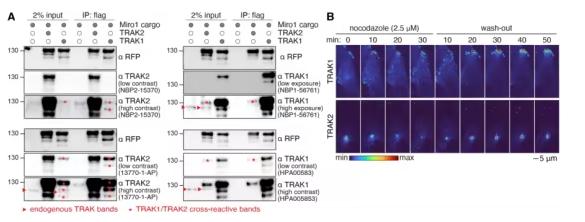
In a new study, Gladkova and colleagues combined synthetic cargo-transport experiments with AlphaFold2-assisted structural analysis to identify a molecular switch that coordinates kinesin- and dynein-driven mitochondrial transport.
They further demonstrated that this switch is regulated by cellular stress.
The characteristics of the mechanism suggest a broader principle that may govern the intracellular distribution of many cellular components transported by microtubule motors.
The researchers combined synthetic cargo-transport assays with AlphaFold2-guided mutational analysis to identify a regulatory helix within the mitochondrial adaptor protein TRAK.
Our Related Proteins
| Cat.No. # | Product Name | Source (Host) | Species | Tag | Protein Length | Price |
|---|---|---|---|---|---|---|
| TRAK-2128S | Recombinant Staphylococcus aureus (strain: CDCPANICU) TRAK protein, His-tagged | E.Coli/Yeast | Staphylococcus | His |

|
|
| TRAK-2147S | Recombinant Staphylococcus aureus (strain: CDCTN147) TRAK protein, His-tagged | E.Coli/Yeast | Staphylococcus | His |
|
|
| TRAK-2170S | Recombinant Staphylococcus aureus (strain: CDCGA672) TRAK protein, His-tagged | E.Coli/Yeast | Staphylococcus | His |
|
|
| TRAK-2215S | Recombinant Staphylococcus aureus (strain: PM79, other: HA-MRSA) TRAK protein, His-tagged | E.Coli/Yeast | Staphylococcus | His |
|
This helix mediates switching between kinesin- and dynein-driven transport.
Differences in the sequence of this helix help explain why two highly similar TRAK isoforms predominantly support mitochondrial transport in opposite directions.
Phosphorylation of the regulatory helix by stress-activated kinases promoted dynein activation and kinesin dissociation.
The findings reveal a molecular mechanism through which intracellular signals can coordinate directional mitochondrial transport.
Disrupted mitochondrial transport has been implicated in Alzheimer's disease, Parkinson's disease, and amyotrophic lateral sclerosis, or ALS.
The structural information provided by this study could therefore contribute to future efforts to identify molecules capable of modulating the TRAK regulatory helix.
8. Natural Sugars, Rather Than Meat Alone, May Have Helped Fuel Human Brain Expansion
DOI: 10.1126/science.aed8437
New research from the University of Sydney and the University of Glasgow suggests that naturally sweet foods such as fruit and honey may have supplied an important source of energy during the evolution of the human brain.
Published in Science, the study used modeling approaches to estimate the glucose requirements of humans and their ancestors over approximately four million years.
The researchers found that before cooking made starch-rich foods such as tubers and grains more digestible, naturally occurring sugars may have provided an important source of glucose needed to support brain growth.
“Raw starch does not release glucose, meaning early humans may have depended on naturally sweet foods for long periods before grains became a routine component of the diet,” said study first author Jennie Brand-Miller, professor of human nutrition at the University of Sydney's Charles Perkins Centre and School of Life and Environmental Sciences.
The findings offer a different perspective on the traditional view that increased meat consumption was a major driver of human evolution.
They may also help explain humans' persistent preference for sweet foods.
“Although animal foods were important, the brain's requirement for glucose has received insufficient attention from anthropologists,” Brand-Miller said.
Over approximately four million years, human brain size increased from around 300 grams in early hominins to about 1,500 grams in modern humans.
As brain size increased, glucose requirements also rose.
Glucose is also required by other tissues, including red blood cells, the kidneys, and reproductive organs.
“The human brain is exceptionally large relative to our body size and requires enormous amounts of energy,” Brand-Miller said. “Glucose is the brain's primary fuel, and during much of human evolution it came from carbohydrates in foods such as fruit and honey.”
9. Candida auris Remodels Its Cell Wall to Manipulate Skin Immunity and Persist in Hair Follicles
DOI: 10.1126/science.adu6688
Candida auris was first identified in 2009 and has since spread rapidly around the world, becoming a major outbreak-associated pathogen in hospitals and long-term care facilities.
Unlike many other Candida species, C. auris can colonize human skin asymptomatically for extended periods and spread through direct contact, potentially causing invasive infection in immunocompromised individuals.
The organism also shows substantial antifungal resistance, with some strains demonstrating reduced susceptibility to all three major classes of antifungal drugs.
Reported mortality in invasive infections can reach 30% to 60%.
Why C. auris can persist so effectively on the skin while its close relative Candida albicans is less successful at long-term skin colonization has remained a major unanswered question.
To investigate the mechanisms underlying C. auris skin colonization, researchers systematically compared the pathogen with C. albicans, a related fungus that colonizes skin less efficiently and is better known as a mucosal commensal.
The study used mouse models, fungal and host genetics, immunological analyses, single-cell RNA sequencing, and volumetric quantitative confocal microscopy.
These approaches allowed researchers to characterize the dynamics and spatial distribution of fungal skin colonization as well as host immune responses, with the goal of identifying microbial and host determinants of persistence and clearance.
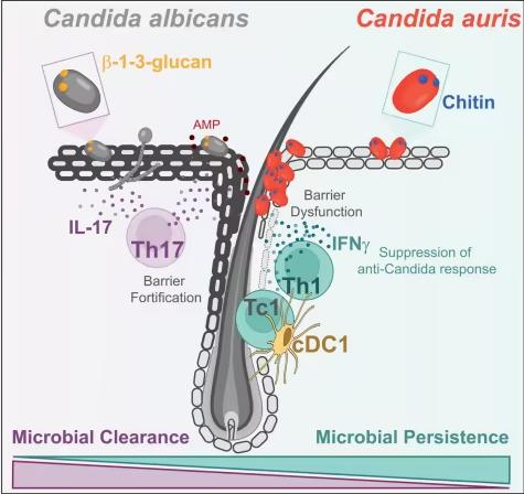
C. auris achieved greater levels and longer persistence of skin colonization in mice than C. albicans. It also displayed direct binding to hair shafts and a pronounced tropism for hair follicles.
As previously reported, C. albicans induced a host-protective type 3/17 cutaneous immune response driven by IL-17A.
By contrast, C. auris triggered an immune response skewed toward type 1 immunity, characterized by expansion of hair-follicle-associated type 1 conventional dendritic cells, or cDC1 cells, type 1 cytotoxic T cells, or Tc1 cells, and Th1 cells, together with increased production of interferon-γ, or IFNγ.
IFNγ signaled directly to hair-follicle keratinocytes and reduced IL-17A-driven gene-expression programs associated with skin-barrier function and antimicrobial defense.
Using mice deficient in specific cytokine-signaling pathways, the researchers established that IFNγ promoted persistent C. auris colonization within the epidermal niche, even though the same cytokine retained its classical host-protective role during deeper skin or bloodstream infection.
When cultured under skin-like conditions—including synthetic sweat, high salt, nutrient-poor medium, and 37°C—C. auris remodeled its cell wall and increased exposure of chitin.
Under the same conditions, C. albicans instead showed increased β-1,3-glucan exposure.
Genetic and pharmacological manipulation of fungal chitin altered skin immune polarization and fungal persistence in vivo, directly linking fungal cell-wall composition with the immune state of the skin and the outcome of colonization.
Our Related Proteins
| Cat.No. # | Product Name | Source (Host) | Species | Tag | Protein Length | Price |
|---|---|---|---|---|---|---|
| IL17A-5633H |
Recombinant Human IL17A protein, His-Avi-tagged, Biotinylated

|
HEK293 | Human | Avi&His | Gly24-Ala155 | |
| IL17A-14153H | Recombinant Human IL17A, GST-tagged | E.coli | Human | GST | 1-155a.a. | |
| IL17A-26H | Recombinant Human Interleukin-17 | E.coli | Human | Non | ||
| Il17a-71M | Recombinant Mouse Interleukin 17A | E.coli | Mouse | Non | ||
| IL17A-12C | Active Recombinant Canine interleukin 17A protein, His tagged | HEK293 | Canine | His | 29-155 aa | |
| Il17a-01M |
Active Recombinant Mouse Il17a Protein, His-Tagged

|
E.coli | Mouse | His |
|
|
| IFNG-510H |
Active Recombinant Human IFNG
|
HEK293 | Human | Non | 24-166 a.a. | |
| IFNG-14081H | Recombinant Human IFNG, His-tagged | E.coli | Human | His | 22-166a.a. | |
| IFNG-4340F | Recombinant Ferret IFNG Protein | Yeast | Ferret | Non | 164aa |
10. A New Antimalarial Strategy Could Turn Future Mosquito Bites Into Natural Immune Boosters
DOI: 10.1126/science.aea7605
Researchers at Australia's Walter and Eliza Hall Institute of Medical Research, or WEHI, have identified a new strategy for preventing malaria—one that could potentially transform future mosquito bites from sources of infection into natural boosters of protective immunity.
The Australian team demonstrated a vaccination strategy in which the immune system is first trained, with the help of drugs, to recognize and attack malaria parasites before they can cause disease.
Subsequent exposure to parasites through natural mosquito bites could then act as an immune booster, reinforcing protection over time.
In this preclinical study published in Science, the researchers developed a new immunization strategy combining mosquito-transmitted malaria parasites with a new class of antimalarial compounds developed by WEHI together with the global biopharmaceutical company MSD.
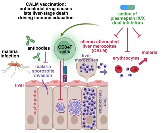
These compounds block parasite development at critical stages of the malaria life cycle, preventing disease while simultaneously triggering a strong immune response capable of providing durable protection.
Subsequent mosquito bites further strengthened the immune response and protective effect.
Associate Professor Justin Boddey, a WEHI laboratory head and corresponding author of the study, said the findings demonstrate how candidate drugs could effectively “pre-activate” the immune system before malaria disease develops.