
Uncategorized Monday, 2026/09/14
As August 2026 draws to a close, what noteworthy studies have been published in Science this month? Here is a roundup of some of the most interesting research highlights.
1. RAD51 Stabilizes Neutrophil Extracellular Traps to Compartmentalize Inflammation
DOI: 10.1126/science.aed9286
Tissue infection triggers local inflammation through the sensing and release of inflammatory mediators. However, the molecular and cellular mechanisms that sustain localized tissue activation while preventing potentially harmful systemic immune activation remain incompletely understood.
Neutrophils are major antimicrobial phagocytes recruited to sites of infection. They protect the host partly by releasing neutrophil extracellular traps, or NETs—web-like structures composed of chromatin and antimicrobial proteins that capture and neutralize pathogens.
Beyond trapping microbes, NETs are also pro-inflammatory, and uncontrolled NET release can contribute to inflammatory disease. NETs possess a distinctive chromatin architecture characterized by large and extensively branched structures. How this organization is maintained and how it influences immune responses have remained unclear.
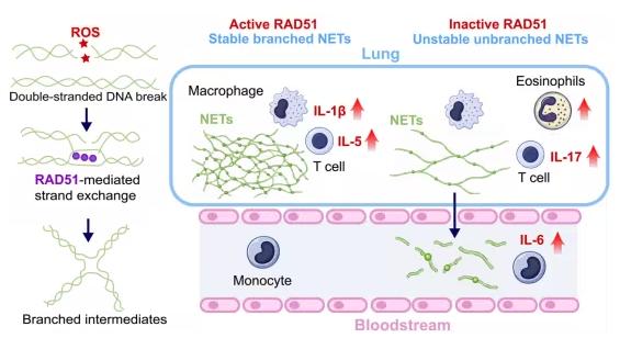
RAD51 promotes homologous DNA recombination repair by generating branched chromatin intermediates known as Holliday junctions. Its role in immunity and neutrophil biology, however, had not been explored.
In a new study, researchers investigated whether RAD51 contributes to the formation of branched NET chromatin fibers and whether alterations in NET branching affect inflammatory signaling. Using imaging, functional assays, inflammatory profiling in experimental models, and analyses of human patient samples, the team examined how RAD51 shapes NET architecture and influences communication between neutrophils and other immune cells, thereby spatially restricting immune activation away from the circulation.
Knockdown of RAD51 in human and mouse neutrophils produced NETs with reduced chromatin branching. These NETs were less stable and more susceptible to degradation by plasma endonucleases. NETs were also sensitive to the branch-targeting resolvase endonucleases GEN1 and RuvC.
Our Related Proteins
| Cat.No. # | Product Name | Source (Host) | Species | Tag | Protein Length | Price |
|---|---|---|---|---|---|---|
| RAD51-128H |
Active Recombinant Full Length Human RAD51, His-tagged

|
E.coli | Human | His | Full L. 1-339 aa | |
| RAD51-134H |
Recombinant Full Length Human RAD51

|
E.coli | Human | Non | Full L. 1-339 aa | |
| RAD51-2155H | Recombinant Human RAD51, GST-tagged | E.coli | Human | GST | 1-242aa | |
| RAD51-02H | Recombinant Human RAD51 Protein, isoform 2 | E.coli | Human |
Different NET-inducing stimuli increased RAD51 expression to varying degrees, producing NETs with different levels of stability.
The researchers initially expected that inhibiting RAD51 in vivo would reduce inflammatory pathology during pulmonary infection with the fungal pathogen Aspergillus fumigatus by accelerating NET clearance. Instead, destabilization of NETs caused pro-inflammatory NET chromatin to rapidly accumulate in the circulation, driving abnormal monocyte activation and interleukin-6, or IL-6, production.
Although lung-derived cytokines such as IL-1β and IL-5 were reduced, dysregulated systemic IL-6 enhanced Th2- and Th17-associated inflammation, promoted eosinophilia, and worsened mucin production and airway obstruction.
Consistent with these findings, patients with aspergillosis showed elevated levels of circulating cell-free DNA, which correlated with IL-6 and the eosinophil chemoattractant eotaxin.
2. Touch and Pain Recruit Different Microarterioles: Neurovascular Coupling Is Not One-Size-Fits-All
DOI: 10.1126/science.aeb5077
The brain continuously adjusts local blood flow in response to increased neuronal activity through a process known as neurovascular coupling.
These vascular responses form the basis of widely used brain-imaging techniques such as functional magnetic resonance imaging, or fMRI, which infer neuronal activity from local changes in blood oxygenation, blood-flow velocity, or blood volume.
Although neurovascular signals are often assumed to reflect neuronal activity in a relatively uniform manner, different sensory experiences recruit distinct neural circuits across different cortical layers. How these differences influence blood-flow regulation in the brain has remained unclear.
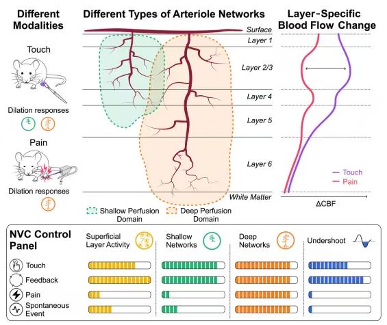
In a new study, researchers hypothesized that neurovascular coupling depends not only on the overall magnitude of neuronal activity, but also on the type of sensory input and how neuronal activity is distributed across cortical layers.
To test this idea, the team compared cortical responses in mice to several forms of neural input, including gentle touch, optogenetic activation of nociceptors, spontaneous activity, and motor-sensory feedback signals.
Using wide-field optical imaging and deep two-photon microscopy, the researchers measured neuronal activity and vascular dynamics across different cortical depths and spatial scales. They then combined the experimental observations with computational simulations to investigate how microarteriole topology shapes blood-flow dynamics.
Touch and nociceptive stimulation produced markedly different blood-flow responses across cortical depths.
Although overall neuronal activity across layers 2 to 5 was broadly similar between the two conditions, nociceptive stimulation reduced the blood-flow response in the superficial cortex, particularly layers 2/3, by more than 50%.
Consistent with this difference, touch produced a larger reduction in deoxyhemoglobin, or HbR, indicating greater blood oxygenation, as well as a more pronounced post-stimulus rebound constriction.
By contrast, blood-flow responses in the deeper layer 6 were similar for touch and pain.
These effects arose because different forms of neuronal activity selectively recruited distinct classes of penetrating microarterioles. Deep microarterioles and their proximal branches dilated across all types of activity, whereas superficial microarterioles dilated only when superficial cortical activity—particularly in layer 1—was sufficiently engaged.
For example, neuropil Ca²⁺ responses in layer 1 were approximately 50% greater during touch than during nociceptive stimulation.
Computational simulations based solely on experimentally measured changes in vessel diameter accurately reproduced the layer-specific perfusion patterns observed in vivo.
3. Capillary Arterialization Drives Collateral Formation After Myocardial Infarction, While Transient VEGF Stimulation Promotes Cardiac Repair
DOI: 10.1126/science.ady3027
Coronary artery disease remains one of the leading causes of death worldwide.
Acute occlusion of a coronary artery deprives downstream heart muscle of oxygen and nutrients, causing myocardial infarction, or MI.
Coronary collateral arteries act as natural bypasses between pre-existing coronary branches. By restoring perfusion to ischemic tissue, these vessels can improve clinical outcomes. However, the cellular origins and molecular mechanisms governing the formation of new collateral arteries have not been fully defined.
Traditional lineage-tracing approaches are limited by inadequate marker specificity and by temporal variability associated with tamoxifen-dependent labeling.
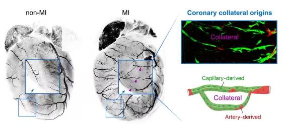
To map the cellular origins of coronary collateral vessels, researchers developed complementary genetic lineage-tracing systems.
These included an “intersectional” strategy designed to reduce false-positive labeling, as well as a tamoxifen-independent system triggered by cell-cell contact that permanently labels mature arterial endothelial cells, or ECs.
The researchers also developed tools capable of simultaneously labeling capillary-derived and artery-derived vessels within the same animal. This allowed them to directly compare the contribution of different EC populations after myocardial infarction and to investigate the signaling pathways regulating the process.
They identified a subset of capillary ECs expressing the arterial marker Cx40, highlighting the specificity limitations of conventional lineage-tracing approaches.
Across multiple independent systems—including intersectional genetics and a synthetic Notch-based method—the researchers found that mature arterial ECs made only a limited contribution to new collateral vessels after myocardial infarction.
Simultaneous lineage tracing within the same heart showed that capillary ECs were the principal building blocks of collateral arteries in both neonatal and adult mice, whereas pre-existing arterial ECs contributed much less.
Selective ablation of capillary-derived collateral vessels impaired tissue repair, increased fibrosis, and worsened cardiac function, confirming their functional necessity.
To enhance collateral formation, the researchers manipulated vascular endothelial growth factor, or VEGF, signaling.
Sustained pathway activation expanded immature artery-like ECs but failed to improve repair.
In contrast, transient delivery of Vegfa using modified mRNA promoted functional collateral formation, improved perfusion, reduced scar formation, and enhanced cardiac function after injury.
Mechanistically, VEGF-A activated the transcription factor YY1, which recruited the chromatin regulator SETD1A, promoted histone H3 lysine 4 trimethylation, or H3K4me3, and induced the arterial regulator HES1, thereby coordinating the transition from capillary to arterial identity.
Our Related Proteins
| Cat.No. # | Product Name | Source (Host) | Species | Tag | Protein Length | Price |
|---|---|---|---|---|---|---|
| VEGFA-210H |
Active Recombinant Human VEGFA protein
|
Insect Cells | Human | Non | 1-209 aa | |
| Vegfa-475M |
Active Recombinant Mouse Vegfa protein(Met1-Arg190)
|
Insect Cells | Mouse | Non | Met1-Arg190 | |
| VEGFA-210HAF647 |
Active Recombinant Human VEGFA Protein, None-tagged, Alexa Fluor 647 conjugated
|
Insect Cells | Human | Non | Met1-Arg209 | |
| Vegfa-38R |
Active Recombinant Rat Vegfa protein, His-tagged, 27-190aa
|
HEK293 | Human | His | 27-190aa | |
| VEGFA-17H | Recombinant Human VEGFA protein | E.coli | Human | Non | 166 | |
| Vegfa-18M | Active Recombinant Mouse Vegfa Protein(N115K) | Yeast | Mouse | Non | 27-190 aa(N115K) |
4. Dystroglycan “Trims” Its Own Glycan Chain: A Phosphorylation-Dephosphorylation Switch Controls Matriglycan Length
DOI: 10.1126/science.adz7427
Dystroglycan, or DG, is a transmembrane receptor widely expressed in tissues including muscle, the nervous system, and the kidneys.
It acts as a molecular bridge between the extracellular matrix and the intracellular cytoskeleton and is essential for maintaining muscle-cell integrity and signaling.
DG function depends heavily on a unique polysaccharide chain attached to its extracellular region called matriglycan.
This long glycan chain extends into the extracellular environment and binds extracellular matrix ligands such as laminin and agrin with high affinity, thereby anchoring cells to the basement membrane.
When matriglycan synthesis is defective or the chain is too short, DG binding is impaired, leading to a group of inherited muscle disorders known as dystroglycanopathies.
These disorders range from relatively mild late-onset myopathies to severe congenital muscular dystrophies such as Walker-Warburg syndrome and may also involve abnormalities of brain development and the eyes.
However, the mechanism controlling matriglycan chain length has remained unclear.
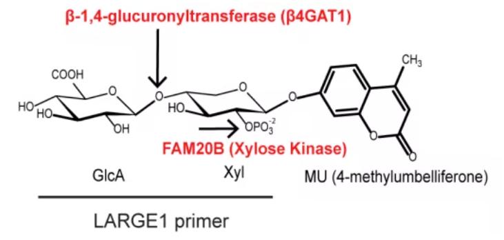
In a new study, Chandel and colleagues found that matriglycan length is regulated by a glycan phosphorylation-dephosphorylation mechanism.
A xylosyl kinase initiates matriglycan synthesis by adding a phosphate group to a xylose residue within the matriglycan primer. When the N-terminal domain of dystroglycan, or DGN, removes this phosphate group, matriglycan can subsequently elongate.
DGN contains a conserved DXDXT/V catalytic motif characteristic of the haloacid dehalogenase family of phosphohydrolases.
Mutation of this site abolished the phosphatase activity of DGN, shortened matriglycan chains, and caused muscle disease in mice.
The findings therefore reveal an unexpected function of dystroglycan itself as a xylose phosphatase involved in controlling matriglycan elongation.
This study provides a new perspective on the mechanisms underlying dystroglycanopathies by showing that DG itself participates in regulating the length of its glycan chain.
The work also suggests potential therapeutic directions.
The conserved DXDXT/V motif within DGN is essential for phosphatase activity. In patients carrying mutations in this region, future gene therapies might potentially aim to correct these sites and restore DG self-regulation.
Base-editing technologies could be particularly relevant for precise correction of certain single-nucleotide mutations.
Because matriglycan initiation requires kinase-mediated phosphorylation whereas elongation requires dephosphorylation by DG itself, the activities of these enzymes could also, in principle, be pharmacologically modulated.
For example, in conditions where insufficient dephosphorylation produces abnormally short glycan chains, compounds that enhance DGN phosphatase activity might be explored. Conversely, defects in initiation could potentially be addressed through modulation of the upstream xylosyl kinase pathway.
5. A Single-Cell, Multi-Organ Atlas Maps Hypertension and Links Disease Mechanisms to Genetic Risk
DOI: 10.1126/science.aea6187
In a new study, researchers developed a comprehensive single-cell atlas of hypertension and hypertension-associated target-organ damage.
The atlas covered three widely used animal models with complementary pathophysiological mechanisms:
- angiotensin II-infused mice, representing neurohumoral mechanisms;
- Dahl salt-sensitive rats, modeling salt-sensitive hypertension; and
- spontaneously hypertensive rats, representing genetically influenced and age-associated hypertension.
The researchers analyzed six organs and tissues, including the hypothalamus, kidney, third-order mesenteric arteries, left ventricle of the heart, middle cerebral artery, and peripheral blood mononuclear cells.
The atlas integrated single-nucleus RNA sequencing, or snRNA-seq, single-cell RNA sequencing, or scRNA-seq, and, in selected samples, snMultiome-seq combining snRNA-seq with single-nucleus ATAC sequencing.
Together, these approaches captured a broad range of transcriptional and epigenetic changes occurring during hypertension progression.
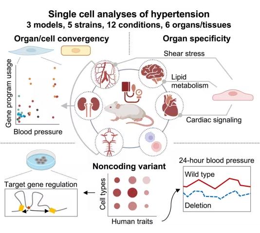
Analysis of the dataset revealed transcriptional changes shared across models and organs, alongside model-specific and tissue-specific adaptive and maladaptive responses.
The researchers identified a conserved vascular smooth-muscle-cell gene-expression program whose activity correlated with blood-pressure levels.
They also identified renal tubular gene programs spanning multiple nephron segments that were associated with kidney injury and blood pressure.
Tissue-specialized endothelial adaptations involved pathways related to lipid handling, shear stress, and cardioprotective signaling.
Coordinated changes across multiple cell types were also observed in specific organs, including the hypothalamus.
The team then integrated these datasets with human genomic information, including genome-wide association study results for blood pressure and eight additional traits related to target-organ injury.
This analysis revealed both model-specific and shared cell type-trait associations, suggesting that different experimental models capture different aspects of the human genetic architecture of hypertension.
The researchers also prioritized a noncoding variant, rs28451064, and used genome editing to demonstrate its influence on blood pressure in vivo.
The variant showed allele-specific regulation of nearby genes including SLC5A3 and RCAN1, potentially involving epigenetic mechanisms such as long-range chromatin interactions.
Our Related Proteins
| Cat.No. # | Product Name | Source (Host) | Species | Tag | Protein Length | Price |
|---|---|---|---|---|---|---|
| RCAN1-088H | Recombinant Human RCAN1 Protein, HIS-tagged | E.coli | Human | His |

|
|
| RCAN1-27792TH | Recombinant Human RCAN1 | E.coli | Human | Non | 1-117 a.a. |
|
| RCAN1-298H | Recombinant Human Regulator of Calcineurin 1 | E.coli | Human | Non | 1-117 a.a. |
|
| RCAN1-4029H | Recombinant Human RCAN1 Protein, GST-tagged | Wheat Germ | Human | GST |
|
|
| RCAN1-4972R | Recombinant Rat RCAN1 Protein | Mammalian Cells | Rat | His |
|
Overall, the study provides a multi-organ and multi-model cellular and molecular resource for hypertension research.
By integrating this atlas with additional datasets and targeted experiments, the researchers identified shared, model-specific, and tissue-specific changes that may contribute to hypertension and its consequences, while also uncovering new mechanisms of gene regulation in the disease.