
Uncategorized Sunday, 2026/03/22
Lymphatic metastasis in breast cancer is not the result of cancer cells acting alone; rather, it is a complex drama jointly staged by tumor cells and their surrounding microenvironment. The systemic interactions among cancer cells, metabolism, and the immune system form the core mechanism that allows metastasis to succeed.
If cancer is a war taking place inside the body, metastasis is often the most troubling chapter. A primary tumor may be removed with surgery, but once cancer cells quietly escape through the lymphatic system and establish new colonies elsewhere in the body, the situation becomes far more difficult to control. In breast cancer, lymph node metastasis is precisely such a dangerous signal—it indicates that cancer cells have learned how to “escape,” and the patient’s prognosis is consequently much worse.
Much of the lethality of breast cancer lies in the word “metastasis.” As the most common cancer among women worldwide, breast cancer accounts for nearly one quarter of all female cancer cases. For most patients, death is ultimately caused not by the original tumor itself, but by the metastatic “offspring” that spread to other parts of the body. Unfortunately, despite the critical importance of lymph node metastasis, scientific understanding of it has long remained limited.
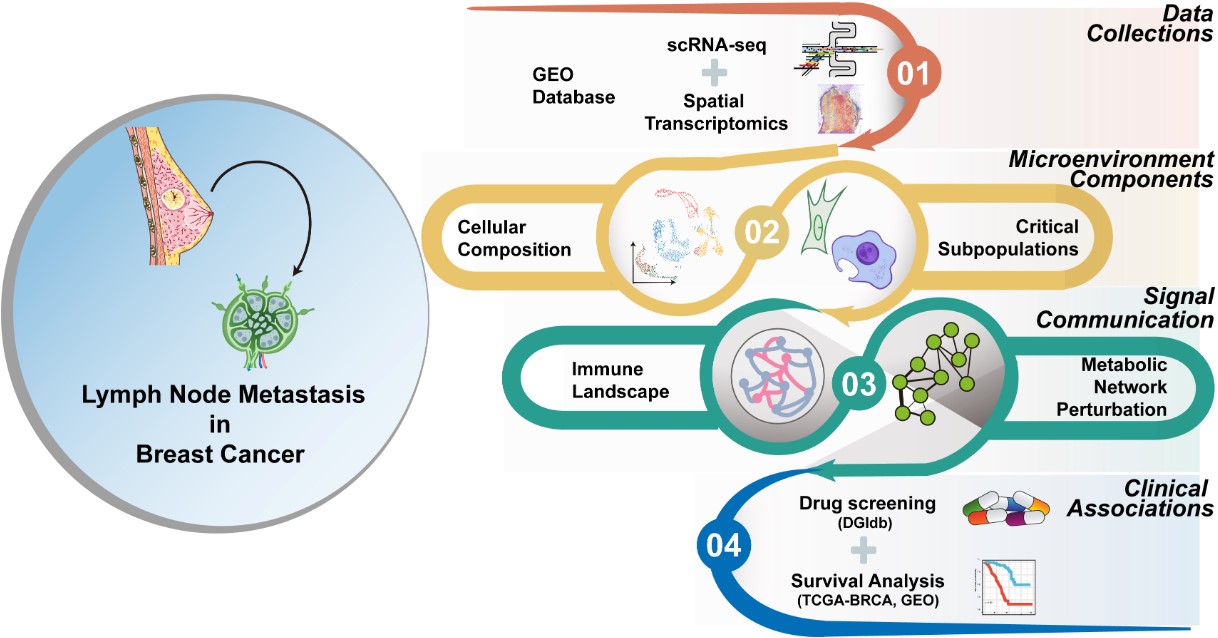
Fig1. Graphic overview of the experimental design in the study.
Recently, a research paper titled “Deciphering the Cellular and Metabolic Landscape of Lymph Node Metastasis in Breast Cancer Using Single-Cell and Spatial Multi-Omics,” published in The American Journal of Pathology, shed new light on this process. Researchers from Nanjing Normal University and other institutions turned powerful microscopic technologies toward the “scene of metastasis,” allowing scientists to see for the first time what cancer cells are actually doing inside lymph nodes.
In this study, the researchers used advanced techniques—single-cell RNA sequencing combined with spatial transcriptomics—to create a high-resolution map of lymphatic metastasis in breast cancer. They analyzed 78 paired samples of primary breast tumors and their corresponding lymph node metastases, examining the characteristics of more than 360,000 individual cells. This was not an ordinary map; it functioned more like a reconstruction of a crime scene, revealing what each cell was doing, whom it was communicating with, and how they were collaborating.
Amid this dense cellular landscape, the researchers identified a special group of cells: early disseminated cancer cells. These cells escape from the primary tumor at a very early stage and lie dormant within lymph nodes. Their danger lies not only in their ability to leave early, but also in their ability to manipulate two key processes.
First is metabolic reprogramming. In oxygen-poor environments, these cells activate glycolysis, essentially creating a “backup power supply” that allows them to survive even under harsh conditions.
Second is immune regulation. These cancer cells interact with nearby immune cells in ways that persuade immune “guards”—which should normally attack tumors—to switch sides and instead provide protection.
This leads to another important discovery. The researchers found that within the metastatic microenvironment there is a complex three-way interaction among cancer cells, macrophages, and lymphocytes. In particular, M2-type macrophages play a key role. These cells secrete signaling molecules such as CCL22 and CXCL12, which both suppress the immune system—preventing cytotoxic T cells from functioning properly—and promote a more aggressive behavior in early disseminated cancer cells.
Our Related Proteins


Evidence from spatial transcriptomics made this interaction even clearer: these “conspiracies” occur in specific regions within lymph nodes that correspond precisely to the frontline areas where tumors advance.
In other words, breast cancer lymphatic metastasis is not the work of cancer cells alone. Instead, it is a coordinated performance involving tumor cells and the surrounding microenvironment. The dynamic interplay among cancer cells, metabolism, and the immune system forms the fundamental mechanism enabling metastasis.
The encouraging news is that once scientists understand this “script,” intervention becomes possible. Using computer-based drug screening, the researchers searched existing medications for candidates that could disrupt this malignant collaboration. They identified four tyrosine kinase inhibitors, including pexidartinib and sunitinib, which are already used to treat other cancers. These drugs target M2 macrophages, and by inhibiting key molecules such as CSF1R, they can potentially interrupt the interaction between cancer cells and macrophages, thereby suppressing lymph node metastasis.
These drugs have already been proven safe in clinical settings and may now have new therapeutic applications. However, the researchers acknowledge that further work is needed—particularly to explore the metabolic vulnerabilities of early disseminated cancer cells and to validate these findings using real clinical data.
In a broader sense, the significance of this study lies in revealing phenomena that were previously invisible. Cancer is often frightening not simply because it exists, but because it is so adept at hiding and adapting. Now, however, scientists have a clearer “treasure map” pointing to its weaknesses and where to look for them. For patients worried about breast cancer metastasis, this represents a promising and hopeful development.
Related Products & Services
- Cell and Gene Therapy
- Immune Checkpoint Proteins
- ADC Target Protein
- PROTAC Targets
- Targets of CAR-T Cell Therapy
- Cancer Drug Targets
- Protein Engineering Services
- Protein Interaction Service
- Protein Expression and Purification Services
- Drug Discovery Screening
- Protein Pathway Profiling
Reference
- Rui Zhu,Guijie Jiang,Jie Shen, et al. Deciphering the Cellular and Metabolic Landscape of Lymph Node Metastasis in Breast Cancer Using Single-Cell and Spatial Multi-Omics. The American Journal of Pathology. DOI:10.1016/j.ajpath.2026.01.002.