
Uncategorized Thursday, 2026/08/06
This study identifies a hypoxia-driven inflammatory and fibrotic program mediated by the hypoxia-inducible factor-1α–phosphoinositide 3-kinase δ–interleukin-13 (HIF-1α–PI3Kδ–IL-13) signaling axis in the skin of patients with chronic graft-versus-host disease (cGVHD).
Chronic graft-versus-host disease (cGVHD) is a major complication following allogeneic hematopoietic stem cell transplantation (allo-HSCT), and skin fibrosis represents one of its most debilitating and treatment-resistant manifestations.
On July 1, 2026, Zhang Xi from Army Medical University and Edmund K. Waller from Emory University published a research article entitled “Hypoxia-driven T cell–macrophage–stromal cross-talk sustains fibrosis in preclinical models of cutaneous chronic graft-versus-host disease” online in Science Translational Medicine. The study revealed a hypoxia-induced inflammatory and fibrotic program in cGVHD skin mediated by the HIF-1α–PI3Kδ–IL-13 signaling pathway.
Using spatial transcriptomics, single-cell RNA sequencing (scRNA-seq), and multiplex immunofluorescence analysis, the researchers demonstrated that HIF-1α stabilization occurs in epidermal cells within hypoxic regions.
HIF-1α stabilization was accompanied by IL-13 production from PI3Kδ-activated T cells and macrophage–epidermal cell interactions, which together promoted fibrosis and the formation of tertiary lymphoid structure (TLS)-like aggregates.
In human epidermal cell lines and induced pluripotent stem cell (iPSC)-derived skin organoids, IL-13 was shown to directly induce HIF-1α expression in keratinocytes under normoxic conditions. Under hypoxic conditions, HIF-1α expression was further enhanced, thereby driving profibrotic signaling pathways.
Pharmacological inhibition of HIF-1α, PI3Kδ, or IL-13 reduced tissue hypoxia, disrupted TLS-like aggregates, and improved disease severity in mouse models of cGVHD.
These findings reveal a targetable regulatory circuit connecting hypoxia, type 2 inflammation, and immune–stromal dysfunction in cGVHD, providing a translational framework for developing therapies against fibrotic cGVHD and other immune-mediated fibrotic disorders.
Our Featured Proteins
| Cat.No. # | Product Name | Source (Host) | Species | Tag | Protein Length | Price |
|---|---|---|---|---|---|---|
| HIF1A-106H | Recombinant Human HIF1A, GST-tagged | Wheat Germ | Human | GST | ||
| HIF1A-145H | Recombinant Human HIF1A protein, His-tagged | E.coli | Human | His | Arg575~Asn826 | |
| HIF1A-198H | Recombinant Human HIF1A protein, GST-tagged | E.coli | Human | GST | 574-799aa | |
| HIF1A-3092H | Recombinant Human HIF1A, His tagged | E.coli | Human | His | 575-826 a.a. | |
| HIF1A-107H |
Recombinant Human HIF1A, His-tagged

|
E.coli | Human | His | 1 to 735aa |

|
| IL13-370H |
Active Recombinant Human IL13

|
CHO | Human | Non | Met 1-Asn 132 | |
| IL13-14149H | Recombinant Human IL13, His-tagged | E.coli | Human | His | 21-146a.a. | |
| IL13-147H | Recombinant Human IL13 protein, hFc-tagged | HEK293 | Human | Fc | Gly21-Asn132 | |
| PIK3CD-29587TH | Recombinant Human PIK3CD | Human | Non | Full L. |
|
|
| PIK3CD-52H | Recombinant Human PIK3CD, GST-tagged | Sf9 Cells | Human | GST |
|
|
| PIK3CD-1727H | Recombinant Human PIK3CD protein, His & T7-tagged | E.coli | Human | His&T7 | Val774~Gln1044 |
|
| IL17A-5633H |
Recombinant Human IL17A protein, His-Avi-tagged, Biotinylated
|
HEK293 | Human | Avi&His | Gly24-Ala155 | |
| IL17A-14153H | Recombinant Human IL17A, GST-tagged | E.coli | Human | GST | 1-155a.a. | |
| IL17A-26H | Recombinant Human Interleukin-17 | E.coli | Human | Non | ||
| Il17a-71M | Recombinant Mouse Interleukin 17A | E.coli | Mouse | Non |
Skin Fibrosis in Chronic Graft-versus-Host Disease: Cellular Interactions and Pathological Mechanisms
Cutaneous chronic graft-versus-host disease (cGVHD) is one of the major causes of morbidity following allogeneic hematopoietic stem cell transplantation (allo-HSCT); however, current therapeutic strategies remain inadequate.
The pathogenesis of cGVHD reflects complex interactions between donor-derived immune cells and host tissues, resulting in chronic inflammation, immune dysregulation, and fibrosis.
Key contributors include:
- T cells;
- B cells;
- autoantibodies;
- macrophages;
- cytokine signaling networks.
Resident skin cells, including keratinocytes and fibroblasts, participate in extracellular matrix (ECM) remodeling after abnormal activation by inflammatory signals and disrupted intercellular communication. These processes lead to excessive ECM production, tissue remodeling, and progressive fibrosis.
However, the cellular interactions that drive cGVHD pathology remain poorly understood.
Hypoxia is increasingly recognized as an important regulator of tissue injury and immune function in conditions such as:
- systemic sclerosis;
- ischemia;
- cancer;
- infection.
In systemic sclerosis, chronic hypoxia caused by vascular dysfunction promotes fibroblast activation and fibrosis through hypoxia-inducible factor-1 (HIF-1)-dependent pathways, accompanied by elevated interleukin-13 (IL-13) levels within a T helper 2 (TH2)-polarized microenvironment.
Under hypoxic conditions, the transcription factor HIF-1α, which coordinates cellular responses to oxygen deprivation, is induced. HIF-1α drives glycolytic reprogramming and influences T-cell activation and lineage differentiation.
Although hypoxia has increasingly been recognized as an immune regulatory factor, its role in cGVHD skin has remained unexplored.
Under persistent hypoxic stress, epidermal cells undergo adaptive and maladaptive changes that may further exacerbate inflammatory and fibrotic processes in cGVHD.
Hypoxia Enhances Macrophage Pathogenic Activity in cGVHD
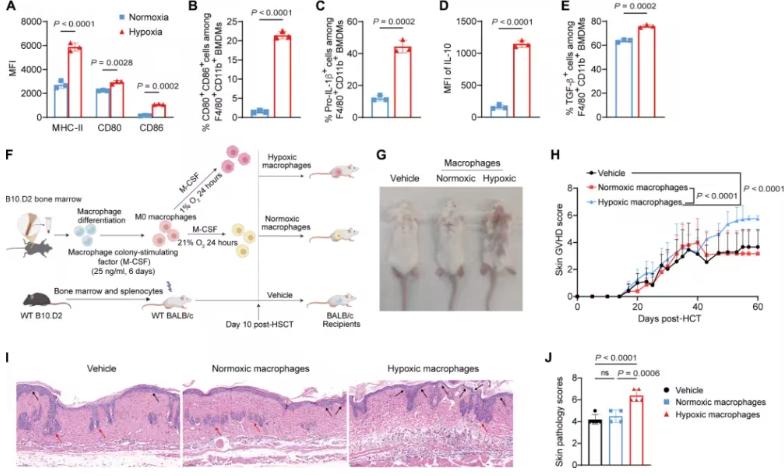
Fig 1. Hypoxia enhances macrophage pathogenic activity in cGVHD (Science Translational Medicine)
Cytokines such as interleukin-17 (IL-17) and IL-13 connect immune activation with hypoxia-driven tissue remodeling.
Primarily produced by TH17 cells, IL-17 amplifies inflammatory responses and stabilizes HIF-1α in stromal cells, maintaining hypoxic conditions through metabolic reprogramming and dysregulated angiogenesis.
Meanwhile, IL-13, secreted by TH2 cells and alternatively activated macrophages, directly promotes fibrosis by stimulating collagen deposition and fibroblast activation. IL-13 also synergizes with hypoxia to enhance HIF-1α signaling.
Noncanonical IL-13 receptor signaling in nonhematopoietic cells involves the phosphoinositide 3-kinase (PI3K) pathway, driving the expression of IL-13-responsive genes such as Tenascin-C, thereby maintaining fibrosis and reinforcing the hypoxic niche.
Although IL-17 and IL-13 have been demonstrated to contribute to experimental cGVHD pathogenesis, how these cytokines coordinate hypoxia-mediated stromal dysfunction within the skin microenvironment remains unclear.
In this study, the authors applied a systems biology approach, integrating:
- spatial transcriptomics;
- single-cell RNA sequencing (scRNA-seq);
- multiplex immunofluorescence analysis;
to dissect the underlying mechanisms of cutaneous cGVHD in both human samples and mouse models.
Related Products & Services
- Cell and Gene Therapy
- Immune Checkpoint Proteins
- ADC Target Protein
- PROTAC Targets
- Targets of CAR-T Cell Therapy
- Cancer Drug Targets
- Protein Engineering Services
- Protein Interaction Service
- Protein Expression and Purification Services
- Drug Discovery Screening
- Protein Pathway Profiling
Reference
- Song, Q., Xu, Y., Wang, X., Wang, W., Feng, Y., Hu, Z., Tian, F., Wang, S., Jankowski, S., Chaudagar, K., Yao, H., Chen, T., Wang, R., Xu, Q., Zhou, Y., Xie, X., He, Y., Wang, Q., Yang, G., . . . Zhang, X. (2026). Hypoxia-driven T cell–macrophage–stromal cross-talk sustains fibrosis in preclinical models of cutaneous chronic graft-versus-host disease. Science Translational Medicine. https://doi.org/adx7264