Uncategorized Tuesday, 2026/05/05
This study used human brain organoids to show that West Nile virus infection can follow two patterns: early clearance or long-term persistence. The virus mainly attacks neurons and astrocytes and triggers the release of multiple inflammatory factors, offering a new perspective for understanding viral encephalitis.
Every summer, mosquito bites are hard to avoid. Most people only feel itchy, put up with it for a while, and then move on. But what you may not know is that a virus called West Nile virus can be hidden in the mouthparts of some mosquitoes. Most people feel little or nothing after infection, but once the virus enters the brain, it can cause deadly encephalitis.
What makes matters worse is that there is currently no vaccine and no specific treatment available worldwide. Even after many patients survive the acute phase, they are left with long-term neurological aftereffects, such as memory problems and unsteady walking. So what exactly happens after the virus enters the brain? Why can some people recover on their own, while others remain ill for a long time?
Recently, a study published in Nature Communications used “mini-brains” grown from human brain tissue to try to answer this question. The results were striking: from the very beginning, infection appeared to follow two different paths.
One Path: Rapid Viral Clearance; the Other: A Long-Term Tug-of-War
The researchers exposed mature brain organoids—roughly equivalent to the human embryonic brain at around 100 days of gestation—to a low dose of West Nile virus. They found that all the organoids could be infected, but their subsequent outcomes were completely different.
In about 56% of the organoids, the viral load reached its peak within four days after infection and then dropped rapidly. Among these, nearly 40%—37.5%—completely cleared the virus within 14 to 28 days. In other words, they managed to eliminate the virus on their own.
Another group of organoids behaved very differently. Their viral peak did not appear until around 11 days after infection, after which they maintained a low level of infection until the end of the experiment without clearing the virus. In other words, they entered a prolonged tug-of-war with the virus.
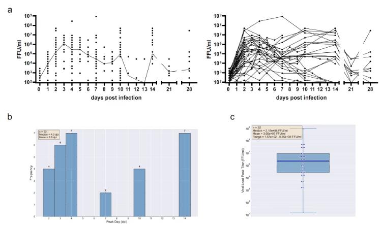
Fig 1. Kinetic characteristics of West Nile virus infection in human brain organoids
The Virus Is Selective About Where It Settles in the Brain
Using fluorescent staining, the researchers observed where the virus was hiding inside the brain organoids. The viral envelope protein appeared only in the outer regions of the organoids, namely the cortex-like structures, and it appeared in small clusters. These areas were packed with two types of cells: neurons and astrocytes. Both cell types were also labeled by the virus, indicating that they were the virus’s main targets.
But one type of cell stood out: microglia. Under normal circumstances, microglia act like the brain’s “patrol guards,” clearing pathogens and cellular debris. In this study, however, microglia never appeared in the virus-clustered regions. Instead, they stayed deeper inside the organoids and were not infected themselves.
In other words, in this brain organoid model, microglia did not actively move to the front line of infection to help.
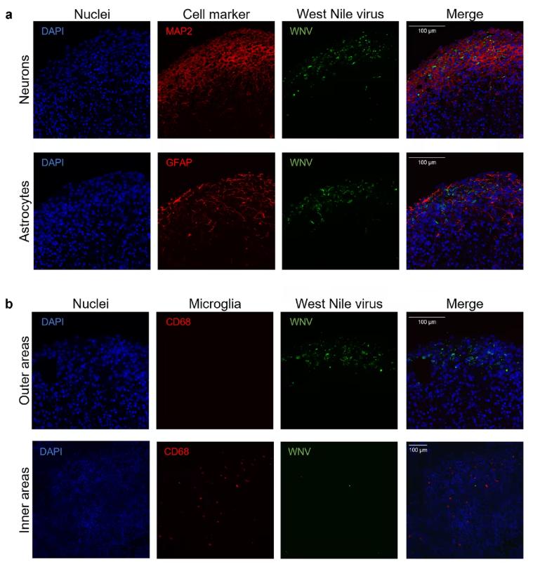
Fig 2. West Nile virus infection localizes to the outer cortex-like regions of human brain organoids
The Timetable of Immune Signaling Molecules: Who Arrives First, and Who Comes Later?
Although microglia did not directly join the battle, the brain organoids still launched their own immune counterattack. The researchers measured various inflammatory signaling molecules in the culture medium from day 2 to day 28 after infection, creating a clear timeline.
The fastest responder was a chemokine called CXCL10, which began to rise by day 2 after infection and remained elevated until day 28. Several other chemokines—CCL17, CCL2, and CX3CL1—rose one after another on day 4. However, CCL17 quickly exited the stage, lasting only until day 7, while CCL2 and CX3CL1 persisted for longer.
Among pro-inflammatory cytokines, TNFα increased from day 4 to day 14; IL-6 and IL-18 appeared from day 7 and day 10, respectively, and both persisted until day 28. Some biomarkers also showed distinctive patterns: for example, IL-1RA appeared only briefly on day 4, while sTREM-1 remained active from day 7 to day 28.
Our Related Proteins


Why Can Some Organoids Clear the Virus? These Signals May Be Involved
The researchers further divided the organoids into two groups: a “rapid clearance group,” in which the viral peak appeared within four days, and a “persistent infection group,” in which the peak appeared around day 14. They also subdivided the organoids according to whether they contained choroid plexus structures, which are tissues capable of producing cerebrospinal fluid.
The results showed that about 64% of the organoids in the rapid clearance group actually cleared the virus. In contrast, none of the organoids in the persistent infection group cleared the virus early on, and only about 15% barely managed to clear it later.
More importantly, the rapid clearance group released significantly higher levels of CXCL10, CX3CL1, IL-1RA, and sTREM-1. These signaling molecules may be key players in helping eliminate the virus. By contrast, a molecule called IL-18 was higher in the persistent infection group, suggesting that it may be associated with the virus “refusing to leave.”
In addition, organoids with choroid plexus structures released more inflammatory signals after infection, indicating that this cerebrospinal-fluid-producing tissue may itself amplify inflammatory responses.

Fig 3. Comparison of inflammatory factor secretion based on organoid morphology and infection progression
What Does This “Mini-Brain” Tell Us?
Using human brain organoids, this study reproduced two different outcomes of West Nile virus encephalitis: in some cases, the brain can quickly clear the virus on its own, while in others, it becomes trapped in long-term low-grade infection. The virus mainly attacks neurons and astrocytes, while microglia do not actively participate in the response. Different infection outcomes correspond to completely different inflammatory signaling profiles.
This model opens a new window into understanding the acute phase of West Nile virus encephalitis and its possible long-term aftereffects. In the future, it could also be used to test antiviral drugs or interventions to see whether “long-term virus-carrying” brains can be shifted into the “rapid viral clearance” category.
Related Products & Services
- RNA Viruses Triggered Signal Pathway
- Diagnostic Antigen
- Diagnostic Antibody
- Protein Engineering Services
- Protein Interaction Service
- Protein Expression and Purification Services
- Drug Discovery Screening
- Protein Pathway Profiling
Reference
- Steffen JF, Widerspick L, Jansen S, Tappe D. A human cerebral organoid model of West Nile virus encephalitis shows innate immunocompetency. Nat Commun. 2026;17(1):2318. Published 2026 Mar 7. doi:10.1038/s41467-026-70281-x