Uncategorized Sunday, 2026/03/22
Only when organoids stop being quirky, one-of-a-kind “only children” and become uniform, standardized “industrial products” can they truly serve as reliable tools for drug screening, disease modeling, and even regenerative medicine.
If you think cell culture simply involves pouring cells into a dish and adding some nutrients, your understanding of biology may still be stuck twenty years in the past. Today, scientists are no longer satisfied with letting cells spread out flat in two dimensions; instead, they want them to “build houses” in three dimensions—growing into structured mini-organs known as organoids. These self-organizing structures can mimic real organ functions and are powerful tools for drug discovery and disease research.
However, reality has been far from ideal. The growth of organoids often resembles a game of chaos: some develop strange shapes, some fail to grow at all, and nine out of ten turn out differently. This “excessive individuality” frustrates researchers. It’s like following the same recipe every time but ending up with completely different dishes—hardly something you can confidently serve.
Even more troubling is the material traditionally used to grow organoids, such as the well-known Matrigel. While it supports cell growth, it is notoriously difficult to work with during the “construction process.” If you want to precisely position cells according to a design, you’re out of luck. At 4°C, it behaves like a liquid, causing cells to sink to the bottom. At 37°C, it quickly solidifies—but the workable window lasts only about two minutes. Before you can properly arrange the cells, the gel sets, trapping them in place or even squeezing them out.
Statistics show that under traditional culture conditions, the coefficient of variation in organoid morphology can exceed 100%. This means that to obtain reliable results, researchers must grow hundreds or thousands of organoids and hope that statistics will compensate for the variability. This is not just an efficiency problem—it’s a crisis of scientific reproducibility. If even a single lab cannot produce consistent batches of “mini intestines,” how can we expect them to reliably predict drug responses in humans?
Cytokines for Organoid Culture


A “Game-Changing” Material
At this critical moment, researchers at the University of California, San Francisco (UCSF) introduced a potential solution. Their study, published in Nature Materials, had a clear goal: to create a material that combines the biological richness of Matrigel with the structural stability of paper—allowing precise cell placement while still supporting self-organization into organoids.
They developed a composite material called the MAGIC matrix. The concept is straightforward: microscopic gel particles made from optically transparent and biologically inert alginate are produced, each roughly the size of a cell. These particles are packed together, and the gaps between them are filled with Matrigel, forming a “sandwich-like” structure that provides both mechanical support and biological nourishment.
At 4°C, the material behaves like wet sand and exhibits shear-thinning properties: when a printing nozzle moves through it, the particles temporarily part, allowing precise deposition of cells. Once the nozzle passes, the particles settle back into place, holding the cells steady. This printing process can last for over two hours, enabling complex patterns.
When the temperature rises to 37°C, the Matrigel in the gaps solidifies, and the material’s mechanical properties become similar to pure Matrigel, creating a favorable environment for cells to continue their self-organization.
The “Golden Rule”: Letting Cells Breathe
But creating the material was only the first step. What truly surprised the researchers was the discovery of a previously overlooked “golden rule”: the quality of organoid growth depends not on how stiff the material is, but on its ability to “relax” under stress.
In simple terms, as cells grow, deform, and expand, the surrounding material must be able to yield and adapt. By adjusting the ratio of microgels to Matrigel, the researchers found that materials capable of rapidly relaxing stress over longer timescales (about one hour) and under large deformations produced intestinal organoids with long, well-formed crypt structures. In contrast, rigid materials that resisted deformation led to stunted and misshapen organoids.
This finding reveals a deeper biological principle: cell self-organization depends not only on chemical signals but also on a physical microenvironment that knows when to “step aside.” The success of the MAGIC matrix lies in its ability to mimic the dynamic mechanical properties of real tissue boundaries—providing both support and flexibility.
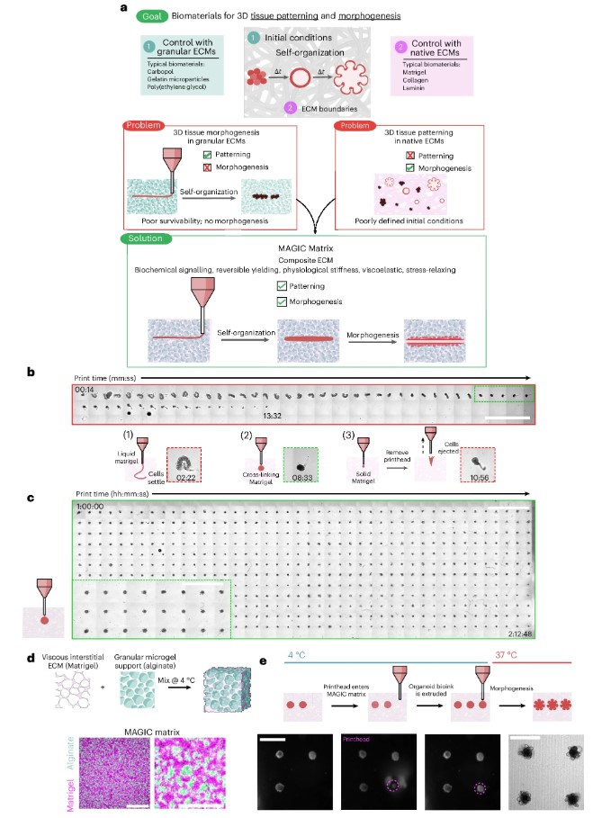
Fig. 1. MAGIC extracellular matrices are embedded bioprinting materials that enable both patterning and morphogenesis of organoids.
From “Handcraft” to Precision Manufacturing
To fully utilize this material, the researchers also developed a piezoelectric-driven bioprinter. This high-precision system can control the volume of extruded material via voltage and even rapidly retract fluid to prevent unwanted trailing.
They dissociated various cell types—including mouse intestinal stem cells, human mammary epithelial cells, vascular endothelial cells, and induced pluripotent stem cell–derived brain organoids—into cell suspensions. These were then printed into the MAGIC matrix as micrometer-scale spheres or tubes, much like writing calligraphy.
The results were remarkable. After printing, the cells quickly self-organized into mature organoids with correct polarity and functional structures. More importantly, they behaved with striking uniformity: hundreds of organoids formed lumens and buds almost simultaneously, with highly consistent size and shape.
In a drug test using a γ-secretase inhibitor, traditional manually seeded organoids required analysis of 135 samples to detect statistical differences. In contrast, printed organoids required only 12 samples, with even clearer results. This represents an order-of-magnitude improvement in efficiency, allowing valuable human biopsy samples to be used far more effectively.
Toward “Organ Factories” and Beyond
The significance of this research goes beyond a new material. It paves the way for organoid research to transition from a “craft-based” approach to precision manufacturing. When organoids become standardized rather than highly variable, they can finally serve as reliable platforms for drug screening, disease modeling, and regenerative medicine.
After all, would you trust drug toxicity tests conducted on a batch of lab mice with wildly different sizes and conditions? The results would hardly be more reliable than drawing lots.
Of course, this is just the beginning. Researchers are now collecting additional data to track organoid development over longer timescales. In the future, they hope to integrate this platform with microfluidics and multicellular co-culture systems to build truly functional “organs-on-chips.”
Perhaps one day, we will witness a tiny, beating heart growing in a dish. And it will all have started with learning how to build cells a well-designed, “well-behaved” three-room home.
Related Products & Services
- Cytokines for Organoid Culture
- Signal Transduction
- Ion Channel Screening Assays
- Protein Pathway Profiling
- Protein Engineering Services
- Protein Interaction Service
- Protein Expression and Purification Services
- Drug Discovery Screening
- Protein Pathway Profiling
Reference
- Graham, A.J., Khoo, M.W.L., Srivastava, V. et al. Stress-relaxing granular bioprinting materials enable complex and uniform organoid self-organization. Nat. Mater. (2026). doi:10.1038/s41563-026-02519-4