RNA Viruses Triggered Signal Pathway
 Full L.
🧪 TBK1-443H
Source: Sf9 Cells
Species: Human
Tag: GST
Conjugation:
Protein Length: 1-729 a.a.
🧪 TBK1-732H
Source: Sf9 Cells
Species: Human
Tag: Non
Conjugation:
Protein Length: 1-729 a.a.

🧪 NFKB2-7052H
Source: Sf9 Cells
Species: Human
Tag: GST
Conjugation:
Protein Length: 1-454 aa

🧪 MAVS-7247H
Source: E.coli
Species: Human
Tag: His
Conjugation:
Protein Length:
🧪 IKBKE-14133H
Source: E.coli
Species: Human
Tag: GST
Conjugation:
Protein Length: C-term-350a.a.
🧪 MAVS-761H
Source: E.coli
Species: Human
Tag: His
Conjugation:
Protein Length: 1-347aa
🧪 NFKB2-1280H
Source: E.coli
Species: Human
Tag: GST
Conjugation:
Protein Length: 600-899 aa
🧪 TRADD-3383H
Source: E.coli
Species: Human
Tag: His
Conjugation:
Protein Length: 1-312 aa
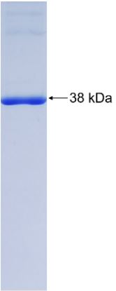
🧪 TRAF3-3384H
Source: E.coli
Species: Human
Tag: GST
Conjugation:
Protein Length: 6-351aa
🧪 TRAF3-3385H
Source: Wheat Germ
Species: Human
Tag: GST
Conjugation:
Protein Length: 1-568 aa
🧪 CASP8-26H
Source: E.coli
Species: Human
Tag: His
Conjugation:
Protein Length: 200-496 a.a.
🧪 IRF3-61H
Source: E.coli
Species: Human
Tag: His&T7
Conjugation:
Protein Length: Met1~Lys360
🧪 TBK1-336H
Source: E.coli
Species: Human
Tag: GST
Conjugation:
Protein Length: 590-729 a.a.
🧪 TBK1-337H
Source: E.coli
Species: Human
Tag: His
Conjugation:
Protein Length: 1-729 a.a.
🧪 FADD-12638H
Source: E.coli
Species: Human
Tag: His
Conjugation:
Protein Length: 1-208a.a.

Background of RNA Viruses Triggered Signal Pathway
What is RNA Viruses Triggered Signal Pathway?
Viruses infect cells to activate innate immune signaling. Innate immunity induce type I interferons production and inflammatory response to clean up viruses from cells. Viruses are divided into DNA viruses and RNA viruses. DNA viruses are recognized by TLRs (Toll-like receptors) and cGAS (cyclic GMP-AMP synthase). On the other way, RNA viruses are recognized by RLRs (RIG-I-like receptors) like RIG-I, MDA5 and LGP2. TLRs and RLRs are both belong to PRRs (pattern recognition receptors). Innate immune signalings triggered by these PRRs lead to transcriptional expression of inflammatory mediators that coordinate the elimination of pathogens and infected cells.
Nucleic acid of RNA viruses is single-stranded RNA (ssRNA) or double-stranded RNA (dsRNA). Many human diseases are caused by RNA viruses include influenza, the common cold, hepatitis C, SARS, Ebola hemorrhoragic fever, West Nile fever and polio etc. It is very important for organism to virus killing and disease prevention by antiviral response. Innate immunity is the most critical process in antiviral response.
RIG-I, MDA5 (melanoma differentiation-associated gene 5), and LGP2 are belong to the RLR (RIG-I-like receptor) family. RLR family proteins are made up of a C-terminal regulatory domain, a central DEAD box helicase/ATPase domain, and two CARDs (N-terminal caspase recruitment domains). They recognize the genomic RNA like dsRNA viruses or dsRNA produced by the replication intermediate of ssRNA viruses. RLRs are localized in the cytoplasm. Virus infection or type I Interferons stimulation can greatly enhance the expression of RLRs.
Both RIG-I and MDA5 are not essential for recognizing Dengue virus and West Nile virus, whereas RIG-I is essential for recognizing hepatitis C virus (HCV). Reovirus is a double-stranded segmented RNA virus, it promotes IFN production mainly through MDA5. However, the lacking of RIG-I or MDA5 both completely reduces the IFN production, indicating that both RIG-I and MDA5 are essential for recognition of reovirus. RNA viruses infect RIG-I-/-MDA5-/-MEFs (mouse embryonic fibroblasts) cannot produce type I IFNs. It means that RIG-I and MDA5 are both essential and sufficient for inducing type I IFN production in response to RNA viruses.
Core Components of RNA Viruses Triggered Signal Pathway
Short dsRNA (up to 1 kb) is recognized by RIG-I, and the type I IFN-inducing activity is greatly enhanced by the presence of its 5’triphosphate. It has been assumed that RIG-I ligand is 5’triphosphate ssRNA produced in vitro. However, RIG-I was not stimulated by chemically synthesized 5’triphosphate ssRNA, it means that the activation of RIG-I needs double-stranded RNA. As for the 19~21mer minimum length dsRNA with a 5’triphosphate end, it can strongly induce type I IFNs. Moreover, synthesized dsRNAs without a 5’phosphate end or with a 5’monophosphate end also activate RIG-I.
The presence of dsRNA is critical for RIG-I recognition. For example, VSV infects cells can produce dsRNA, IFN-b inducing activity will reduce when the produced dsRNA was disrupted. VSV-infected cells generate DI (defective interfering) particles, the size of DI particles are approximately 2.2 kb. Therefore, the dsRNA fragments produced by VSV infection maybe derive from DI particles.
Differ from RIG-I, MDA5 detects long dsRNA (more than 2 kb) like poly I:C. MDA5-/-mice show stongly reduced production of type I IFNs but not IL-12 in response to poly I:C stimulation in vivo. Poly I:C become a RIG-I ligand from an MDA5 ligand by cutting down the length of the poly I:C by dsRNA-specific nuclease. It is indicated that long, but not short, dsRNA is recognized by MDA5.
The other member of RLR family LGP2 lacks a CARD domain. In vitro studies suggested that LGP2 is a negative regulator of RIG-I and MDA5 signaling. LGP2 can isolate dsRNA and inhibit RIG-I conformational changes. However, the in vivo studies of LGP2-/-mice indicated that LGP2 positively regulates both RIG-I and MDA5 mediated production of type I IFNs. Nevertheless, LGP2 is dispensable for type I IFN production after stimulation by synthetic RNAs.
C-terminal regulatory domain (RD) of RLRs family is used for binding to dsRNAs. Recent studies have shown that RIG-I and LGP2’s RD have large basic surface to form RNA-binding loops. The termini of dsRNA bind to the RNA-binding loop. However, MDA5 C-terminal RD is different from RIG-I and LGP2. MDA5 RD also has a large basic surface, but it will not from RNA-binding loop. Therefore, the activity of RNA-binding of MDA5 is much weaker than RIG-I and LGP2. DExD/H helicase domain of RLRs catalyze ATP to induce type I IFN production.

Fig1. Structure‐based model of RIG‐I activation. (Daniel Thoresen, 2021)
RNA Viruses Triggered IPS-1 Downstream Signal Pathway
RLRs adaptor IPS-1 (IFN-b-promoter stimulator 1), also known as MAVS or VISA, has N-terminal CARD-domain. The CARD-domain interacts with CARDs of RLRs for triggering signaling axis. IPS-1 is located on the mitochondrial membrane. When it was cut from mitochondria by HCV NS3/4A protease, RLR signaling activity would disappear. NOD9 is reported as an inhibitor of IPS-1 which belong to NLR family member and is located on mitochondria, but recent studies showed that NOD9 is responsible for reactive oxygen generation. Therefore, the relationship of NOD9 and RLR signaling needs further studies. IPS-1 activates downstream proteins TRAF3 and TRADD. TRAF3 activates IKK-i/TBK1 (TANK-binding kinase 1), then TBK1 dimerize and pass signaling to downstream protein IRF3 (Interferon regulatory Factor 3) and IRF7 (Interferon regulatory Factor 7). IRF3 and IRF7 are phosphorylated and enter nucleus to induce type I interferons production. Another signaling is activated by TRADD and FADD. TRADD and FADD transmit signal to result in the Cleavage of caspase-8/-10. This process activates NF-κB to induce cytokines production. Both TRIF and IPS-1 signaling pathways are regulating the IFN production and the expression of IFN-induced genes.
Case Study
Case Study 1: Recombinant Human MAVS (MAVS-7247H)
The innate immune response (IIR) is a coordinated intracellular signaling network activated by the presence of pathogen-associated molecular patterns that limits pathogen spread and induces adaptive immunity. Although the precise temporal activation of the various arms of the IIR is a critical factor in the outcome of a disease, currently there are no quantitative multiplex methods for its measurement. In this study, the researchers investigate the temporal activation pattern of the IIR in response to intracellular double-stranded RNA stimulation using a quantitative 10-plex stable isotope dilution-selected reaction monitoring-MS assay. We were able to observe rapid activation of both NF-κB and IRF3 signaling arms, with IRF3 demonstrating a transient response, whereas NF-κB underwent a delayed secondary amplification phase. Later in the time course of stimulation, the nuclear IκBα pool is repopulated first prior to its cytoplasmic accumulation. Examination of the IRF3 pathway components shows that double-stranded RNA induces initial consumption of the RIG-I PRR and the IRF3 kinase (TBK1). Stable isotope dilution-selected reaction monitoring-MS measurements after siRNA-mediated IRF3 or RelA knockdown suggests that a low nuclear threshold of NF-κB is required for inducing target gene expression, and that there is cross-inhibition of the NF-κB and IRF3 signaling arms.

Fig1. SID-SRM-MS analysis profiles of cytoplasmic RIG-I, MAVS, IKKα, TBK1, and IKKγ. (Yingxin Zhao, 2013)
Case Study 2: Recombinant Human TRAF3 protein
Cardiac infarction is a dynamic, nonlinear and unpredictable course of disease, and who die of acute myocardial infarction, and coronary thrombosis. TRAF3 provide novel targets for the clinical prevention and treatment for tumors, viral infection, and so on.The researchers investigated the mechanisms of TRAF3 gene, which plays a possible role in cardiac infarction and contributes to the pathogenesis of cardiac infarction-induced oxidative stress. Serum samples of patients with cardiac infarction and normal healthy volunteers were obtained from the 920 Hospital of PLA joint service support force. C57BL/6 mice were ligated and H9C2 cells were induced with 1% O2,5%CO2 and 94% N2. The results showed that over-expression of TRAF3 heightened ROS-induced oxidative stress in vitro model of cardiac infarction. Then, TRAF3 recombinant protein could promote oxidative stress and aggravated cardiac infarction in mice model. Over-expression of TRAF3 induced ULK1 protein expression and reduced ULK1 ubiquitination in vitro model. The activation of ULK1 reduced the effects of TRAF3 on oxidative stress in vitro model of cardiac infarction.

Fig2. The expression of Flag-ULK1 and HA-TRAF3 mutants were analyzed. (Shaobing Zhu, 2022)
Case Study 3: Recombinant Human IRF3
The transcription factor interferon regulatory factor 3 (IRF3) is essential for virus infection-triggered induction of type I interferons (IFN-I) and innate immune responses. IRF3 activity is tightly regulated by conventional posttranslational modifications (PTMs) such as phosphorylation and ubiquitination. Here, the researchers identify an unconventional PTM of IRF3 that directly inhibits its transcriptional activity and attenuates antiviral immune response. They performed an RNA interference screen and found that lysine acetyltransferase 8 (KAT8), which is ubiquitously expressed in immune cells (particularly in macrophages), selectively inhibits RNA and DNA virus-triggered IFN-I production in macrophages and dendritic cells. KAT8 deficiency protects mice from viral challenge by enhancing IFN-I production. Mechanistically, KAT8 directly interacts with IRF3 and mediates IRF3 acetylation at lysine 359 via its MYST domain. KAT8 inhibits IRF3 recruitment to IFN-I gene promoters and decreases the transcriptional activity of IRF3.

Fig3. Immunoblot analysis of IRF3 dimerization. (Wanwan Huai, 2019)
Related Products
When infected with RNA viruses, RNA receptors in host cells, such as RIG-I and MDA5, recognize viral RNA and activate TBK1 and IRF3 via MAVS (mitochondrial antiviral signaling proteins), which in turn promote the expression of IFN and inflammatory cytokines. Creative BioMart can provide a list of core protein products of RLR (RIG-I like receptor) Signaling Pathway or other related pathways to help you with your researches. Please feel free to contact us if you’re interested.

