Histone deacetylation is achieved by the catalysis of histone deacetylase (HDAC) that mediates chromatin remodeling and gene expression regulation. In addition to histones, nicotinamide adenine dinucleotide (NAD)-independent class II HDAC6 or the NAD-dependent deacetylase sirtuin 2 (SIRT2) also binds to and deacetylates α-tubulin, which is a hallmark of long-lived stable microtubules. Post-translational modifications (PTMs) occurring on microtubules are implicated in the regulation of microtubule properties and functions. HDAC6 located mainly in the cytoplasm has been an intensively studied therapeutic target due to its implications in neurodegenerative disorders, depressive behaviors and immune activities. Histone hyperacetylation induced by HDAC inhibitors is associated with gene expression, cell differentiation, cell-cycle arrest, and cell death. Additionally, HDAC inhibitors have been recommended for the treatment of cancers and neurodegenerative disorders. For example, small molecule “tubacin” that inhibits α-tubulin deacetylation in mammalian cells does not affect the stability of microtubules but decreases cell motility. The elucidation of the function for histone deacetylation is necessary to better understand the physiological targets of HDACs and the mechanism by which HDAC inhibitors mediate their spectrum of phenotypic effects.
Creative BioMart offers a panel of different cell-based histone & tubulin deacetylation assays for early drug discovery. Based on Western blot technology, a multidimensional, high-throughput, cell-based screening of HDAC inhibitors can be performed in a suite of tumor cell lines such as A549 and Hela, as well as in any cell line of your choice. The ability to suppress the histone & tubulin deacetylation of a small-molecule chemical genetic modifier is investigated, which allows further classification of the library molecules into functionally related groups.
Procedure of Cell-based Histone & Tubulin Deacetylation Assays at Creative BioMart
Two groups of state-specific antibodies, anti-acetylated α-tubulin and anti-α-tubulin, or anti-acetylated histone and anti-histone antibodies are exploited in Western blot assays to distinguish between small molecules capable of inducing α-tubulin acetylation and histone acetylation. First, cell lines are treated with HDAC inhibitors or DMSO (dimethylsulphoxide) as control in the presence of buffer for a few hours. The whole cell lysates of the treated cells are then subject to Western blot analyses with either group of antibodies. The specific signals of bands of interest are quantified and calculated by Gel-Pro Analyzer. In this assay, cultured primary cell, cell line, or tissue homogenate can be assayed for HDAC activity if the appropriate dose of HDAC specific inhibitor is used.
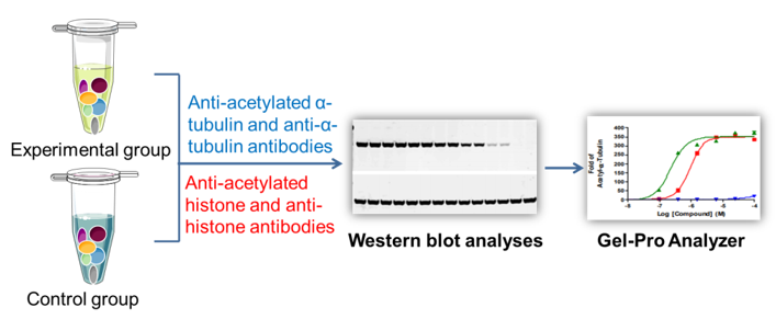
Figure 1. Workflow of cell-based histone & tubulin deacetylation assay
A broad range of targets, technologies, and/or applications at Creative BioMart can be leveraged or adapted to meet your specific needs. With flexible solutions, project management support, prompt communication, on-time data delivery and budget reduction, we are devoted to helping you achieve your project goals with short turnaround time. For more detailed information, please feel free to contact us.
Reference
1. Miyake, Y.; et al. Structural insights into HDAC6 tubulin deacetylation and its selective inhibition. Nat Chem Biol. 2016, 12: 748–754.
USA
Enter your email here to subscribe.
Follow us on

Easy access to products and services you need from our library via powerful searching tools