
Principle and Protocol of Coomassie Brilliant Blue
Coomassie brilliant blue staining method can reach the detection level of microgram level, and the linear dynamic correlation range between staining degree and protein content is wide, which is suitable for quantitative analysis. This method has been widely used due to its low cost, convenient use and good compatibility with the subsequent mass spectrometry identification technology. Coomassie brilliant blue staining has high sensitivity, and 0.1% can be detected in polyacrylamide gel μ G protein. However, for the micro expressed proteins, the detection level of microgram level makes it miss a lot of low abundance proteins. Therefore, other more sensitive staining methods are needed for the detection of low abundance proteins. Coomassie brilliant blue is closely bound to some paper media, so it cannot be used for dyeing filter paper, cellulose acetate film and protein imprinting (on nitrocellulose paper). In this case, the protein is usually denatured by soaking it in 10% trichloroacetic acid, and then dyed with dyes that do not strongly dye the medium, such as bromophenol blue and amino black.
Coomassie brilliant blue R-250 (CBB) is the most commonly used dye in Coomassie brilliant blue dyeing method. Usually, 0.1% or 0.25% Coomassie brilliant blue solution is prepared with methanol: water: glacial acetic acid (volume ratio: 40:50:10) as the dyeing solution. This acetic acid methanol solution denatures the protein and fixes it in the gel to prevent the protein from diffusing in the gel during the dyeing process, which usually takes 2h. The decolorizing solution is the same acetic acid methanol mixture, but does not contain dye, and the decolorization usually needs to be carried out overnight. In order to reduce the irritation and toxicity of acetic acid and methanol, phosphoric acid and ethanol can also be used instead. Methanol and ethanol play a fixed role in dyeing, fixing proteins in gel or preventing their diffusion in gel, and also removing substances left in the electrophoresis process that interfere with the dyeing process, such as detergent, reducing agent, buffer and other components. The role of acetic acid not only helps to fix proteins, but also maintains the pH of the dye solution to facilitate the binding of Coomassie Brilliant Blue with proteins. The existence of them is conducive to obtaining images with clear background and high signal-to-noise ratio.
1. Main Instruments and Equipment
Micropipette, decolorization shaker, balance.
2. Experimental Materials
PAGE gel of protein sample.
3. Main reagents
Methanol, glacial acetic acid, double distilled water, Coomassie brilliant blue R-250, TCA (trichloroacetic acid) or hydrochloric acid, Coomassie brilliant blue G-250.
1. Classic Coomassie Brilliant Blue R-250 Staining
(1) Fixation: After electrophoresis, transfer the gel into a clean glass or plastic container. Add 20% TCA solution, the solution must be free of glue, and fix for 1h.
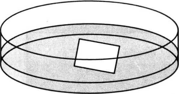
(2) Staining: transfer the gel into 0.1% Coomassie Brilliant Blue staining solution (0.1g Coomassie Brilliant Blue R-250 is dissolved in 40mL methanol, 50mL deionized water and 10mL glacial acetic acid), decolorize and shake the shaking table for dyeing 2h*1.
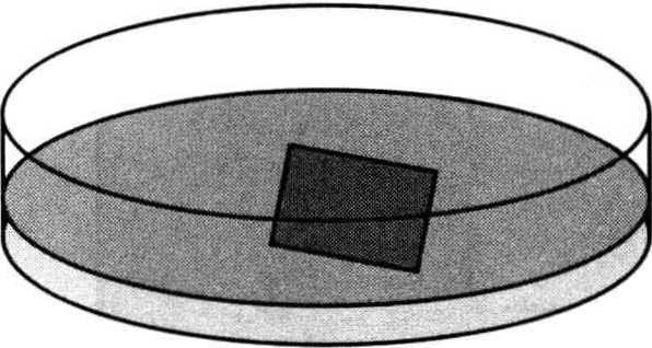
(3) Decolorization: remove the dye solution (it can be reused for 20-40 times after collection and storage), add the decolorization solution (excluding 40% methanol and 10% acetic acid of Coomassie Brilliant Blue R-250), shake at room temperature for decolorization until the background is transparent and clear*2.
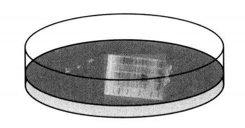
(4) Strengthening: transfer the gel into 1% acetic acid and shake it overnight.
(5) Cleaning: add deionized water for cleaning, and shake the decolorization shaker for 30min.
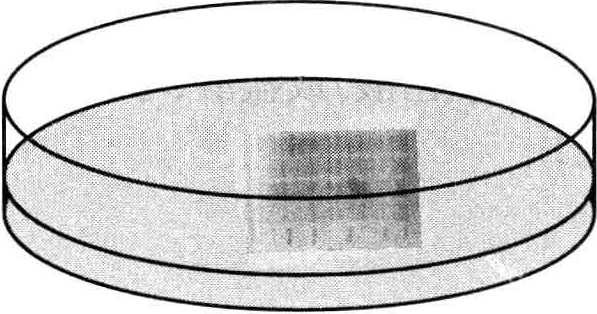
(6) Scan the gel image.
2.Improved Coomassie Brilliant Blue G-250 Staining
(1) After electrophoresis, take out the gel and immerse it in 5 mmol/L hydrochloric acid solution, and shake it for 1h.
(2) Discard the hydrochloric acid solution and repeat step (1).
(3) Put glue in dye solution*3, dye for 2-16h, and shake continuously.
(4) Discard the dye solution and decolorize the glue in 1mmol/L hydrochloric acid solution for several hours (change the solution 3-5 times).
(5) Observe and photograph the gel, and store the gel in 1mmol/L hydrochloric acid solution.
- Sometimes, in order to save time, you can directly dye and decolor without fixing and strengthening. Generally, dye for 20 min to 2 h, and then decolorize until the strips are clear and the background is transparent.
- Using heated dye solution or decolorizing solution (50-60 °C) can shorten the time of dyeing or decolorization.
- The improved method is better for SDS-PAGE and conventional PAGE, but worse for IEF gel dyeing.
*1 The dyeing time can be changed flexibly, and the dyeing time can be shortened at 37 °C.
*2 Be careful not to decolorize excessively.
*3 Take 1mL 1mol/L hydrochloric acid solution, 15mL 1g/L Coomassie Brilliant Blue G-250 solution, mix with water to 1L, stir and mix well.