Principle and Protocol of Silver Stain
Silver staining is an important dyeing method, which is widely used because of its low cost, safe reagents, fast and simple, and high sensitivity. Protein silver staining is a more sensitive method than Coomassie brilliant blue staining. It indicates the protein zone by reducing silver ions (Ag+) to metallic silver on the protein to form black. Silver staining can be carried out directly or after coomassie brilliant blue staining, so that the main protein bands of the gel can be identified by coomassie brilliant blue staining, while silver staining can be detected in the small protein bands that cannot be detected by coomassie brilliant blue staining. The sensitivity of silver staining is 100 times higher than that of Coomassie brilliant blue staining, and it can detect proteins lower than 1ng. However, compared with Coomassie Brilliant Blue staining, silver staining steps are complex and cumbersome, and the coverage of linear coloring dynamics is narrow, leading to the inaccuracy of protein difference display. At the same time, due to the existence of free silver ions and related reagents, it brings some practical difficulties to the subsequent analysis and identification.
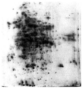
The silver staining of protein bands is based on the combination of various groups (such as carbonyl, carboxyl, etc.) in the protein with silver, which can detect nanogram protein bands. The principle is to use formaldehyde to reduce silver nitrate (silver ion) on the protein belt to metallic silver under specific conditions, so that silver particles can be deposited on the surface of the protein to develop color. At the same time, the silver complexing agent sodium thiosulfate prevents free silver ions from being reduced to metallic silver, which can reduce non-specific staining and background color. The degree of staining is related to some special groups in the protein. Proteins without or with few cysteine residues sometimes show negative staining. Silver staining is widely used in two-dimensional electrophoresis gel analysis and vertical gel for determination of extremely low protein content due to its high sensitivity, which can dye spots below 1ng/protein on the gel.
1. Main Instruments and Equipment
Micropipette, decolorization shaker, balance.
2. Experimental Materials
PAGE gel of protein sample.
3. Main reagents
Silver staining reagents (all are ready to use):
Stationary solution (10% acetic acid, 40% methanol): 200 mL methanol, 50 mL glacial acetic acid, 250 mL double distilled water.
Sensitization solution: methanol 75mL, glutaraldehyde (25%) 1.25mL, sodium thiosulfate (5%) 10mL, sodium acetate 17g, add double distilled water to 250mL and mix well.
Dye solution: dissolve 1g AgNO3 in 1000mL double distilled water, and add 1.5mL 37% formaldehyde solution before use.
Developer: 6.25g sodium carbonate, 0.05mL formaldehyde (37%), add double distilled water to 250mL.
Termination solution: EDTA-Na2 · 2H2O 3.65g, add double distilled water to 250mL.
Reagents required to improve silver staining method:
Stationary solution (10% acetic acid): 50 mL of glacial acetic acid, 450 mL of double distilled water.
Dye solution: dissolve 1g AgNO3 in 1000mL double distilled water, and add 1.5mL 37% formaldehyde solution before use.
Developer: dissolve 30g Na2CO3 in 1000mL double distilled water, cool to 4°C. Before use, add 1.5mL 37% formaldehyde solution, 0.1 mol/L Na2S2O3·5H2O 150 μL; (0.1 mol/L Na2S2O3·5H2O: dissolve 2.48g Na2S2O3·5H2O in 100mL double distilled water).
Termination solution: the same as fixed solution.
Experimental Methods
1. Classic Silver Dyeing
(1) Fixation: put a glass plate with gel into a plate containing 500mL fixative *1, shake the decolorizing shaker for 30min, and wash it with double distilled water for three times.
(2) Sensitization: add sensitizing solution, shake the decolorization shaker for 30min, and wash with double distilled water for 3 times*2.
(3) Silver staining: put the glass plate into a plate containing 500mL of dye solution, shake it on a horizontal shaker for 20min and wash it twice*3 with double distilled water.
(4) Developing: add developing solution, shake the decolorization shaker for 2-3min, and wash it with double distilled water for 3 times.
(5) Stopping display: add stop liquid, and shake the decolorization shaker for 10min.
2. Improved Silver Dyeing
(1) Fixation: gently separate the glass plate with a knife, and put a glass plate with gel into a 500ml container Put the stationary liquid in a plate on a horizontal shaker and shake it for 30min or until the dye band completely disappears.
(2)Washing: pour the fixative back into the beaker, and then wash it with double distilled water for 3 times, 2min each time.
(3) Dyeing: put the glass plate into a plate containing 1L dye solution*4, and shake it on a horizontal shaker for 20 minutes.
(4 )Development: put the glass plate in the precooled double distilled water for a short time (no more than 10s), and quickly put it into the precooled (4°C) developing solution, and develop the color for 3-5 minutes or until all the bands appear*5.
(5) Stop: When clear strips appear, add 500mL of previously used fixative and gently shake for 3min to stop development.
(6) Wash the gel twice with double distilled water for 2min each time.
(7) Dry and scan.

- The level of silver staining for protein detection is about 100ng. Compared with Coomassie brilliant blue staining, the sample amount of silver staining is less, otherwise it is difficult to obtain clear electrophoresis resolution.
- The temperature of silver dye developer should not be too high, and it should be about 10°C. If it is high, it is easy to develop excessively, if the background is deep, it will take a long time to develop. 3. If the fixing time is long, add a step of water washing for 30min to prevent the glue from being too brittle and breaking.
- Formaldehyde shall be added before use.
- It is better to prepare more than one color developing solution. When the solution becomes turbid during the first color development, change a color developing solution, and the color development is clear.
- The utensils used should be clean and do not touch directly with hands to avoid contamination by foreign proteins.
- Washing after silver immersion and before color development is very important, and the time should not be too long or too many times, otherwise the color may not be dyed. However, insufficient washing may lead to a deep background. Generally, it is washed three times with water, about half a minute each time.
*1 Clean utensils should be used to avoid direct contact with gel with hands. Disposable gloves or powder free latex gloves must be worn.
*2 After sensitization, the cleaning times can be increased to appropriately reduce the background color.
*3 The purity of water in the washing process has a great influence on the dyeing result, so it is recommended to use sterile deionized water.
*4 The silver dye solution and color developing solution need to be precooled.
*5 The color development process is very fast. Pay attention to the time to avoid excessive dyeing


