
PVALB
-
Official Full Name
Pvalb parvalbumin
-
Synonyms
PVALB; parvalbumin; parvalbumin alpha; PV; Pva; Parv;
- Recombinant Proteins
- Cell & Tissue Lysates
- Protein Pre-coupled Magnetic Beads
- Chicken
- Human
- Mouse
- Rat
- E.coli
- HEK293
- HEK293T
- Mammalian Cell
- C
- His
- GST
- His (Fc)
- Avi
- SUMO
- Myc
- DDK
- N/A
- N
- Background
- Quality Guarantee
- Case Study
- Involved Pathway
- Protein Function
- Interacting Protein
- PVALB Related Research Area
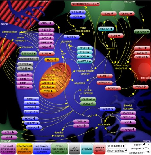
Fig1. The interplay of cell types and pathways in sporadic ALS (SALS) pathology.
What is PVALB protein?
PVALB (parvalbumin) gene is a protein coding gene which situated on the long arm of chromosome 22 at locus 22q12. Parvalbumin alpha, also known PVALB, belongs to a larger group of EF hand proteins. It is a high affinity calcium ion-binding protein that is structurally and functionally similar to calmodulin and troponin C. Parvalbumin is expressed in a specific population of GABAergic interneurones which are thought to play a role in maintaining the balance between excitation and inhibition in the cortex as well as the hippocampus. The PVALB protein is consisted of 110 amino acids and its molecular mass is approximately 12.1 kDa.
What is the function of PVALB protein?
PVALB is a small molecule calcium-binding protein that is mainly found in neurons of the central nervous system and plays a key role in the function and regulation of neurons. PVALB regulates neuronal excitability by binding and regulating the concentration of free calcium ions in cells. When PVALB binds to calcium ions, neuronal excitability decreases. When PVALB releases calcium ions, neuronal excitability increases. PVALB interacts with some sodium ion channels, making it also important in synaptic plasticity.
PVALB Related Signaling Pathway
PVALB protein binds to calcium ion with high affinity and can participate in signal transduction, muscle contraction and apoptosis. It can also influence transmitter release in presynaptic membrane and ion channel activity in postsynaptic membrane, thereby regulating the efficiency and pattern of synaptic transmission. It releases GABA in presynaptic neurons to inhibit neuronal excitability and is involved in regulating the synchrony and rhythm of neuronal networks.
PVALB Related Diseases
The abnormal expression of PVALB protein is closely related to the occurrence and development of neurological diseases such as epilepsy, Parkinson's disease and schizophrenia. In addition, abnormal PVALB function may be associated with brain injury, psychiatric disorders such as anxiety and depression.
Bioapplications of PVALB
Because PVALB plays an important role in a variety of diseases, it is also seen as a potential drug target. Researchers are exploring small molecule inhibitors or agonists that target PVALB to treat diseases such as Alzheimer's disease and epilepsy. PVALB is also used in the field of biotechnology, such as for the production of recombinant proteins, the development of new biological materials, and so on.
High Purity
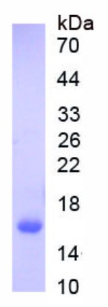
Fig1. SDS-PAGE (PVALB-2644H) (PROTOCOL for western blot)
.
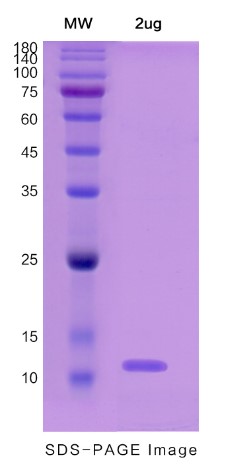
Fig2. SDS-PAGE (PVALB-6118H) (PROTOCOL for western blot)
Case study 1: Janine Wörthmüller, 2018
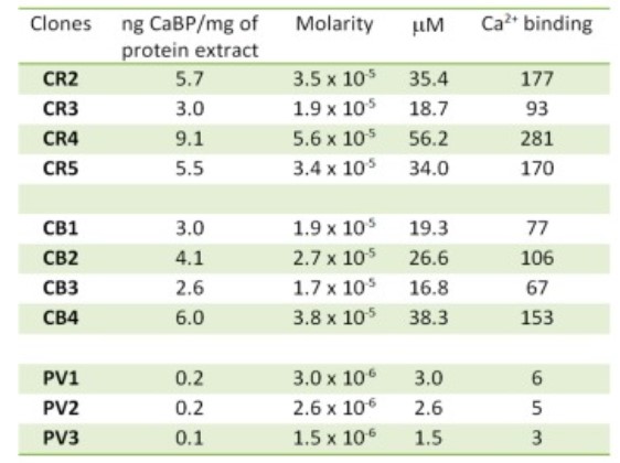
Calretinin (CR; CALB2) belonging to the family of EF-hand Ca2+-binding proteins (CaBP) is widely used as a positive marker for the identification of human malignant mesothelioma (MM). No studies have yet investigated whether other closely related CaBPs might serve as substitutes for CR's functions(s) in MM cells.
Genetically modified MM cell lines with medium (MSTO-211H and ZL5) or low (SPC111) endogenous CR expression levels were generated that overexpress either CR's closest homologue calbindin-D28k (CB) or parvalbumin (PV). After lentiviral shCALB2-mediated CR downregulation, in both MSTO-211H and ZL5 cells expressing CB or PV, the CR deficiency-mediated increase in cell death was not prevented by CB or PV. In conclusion, CR has a likely unique role in MM that cannot be substituted by "similar" CaBPs.
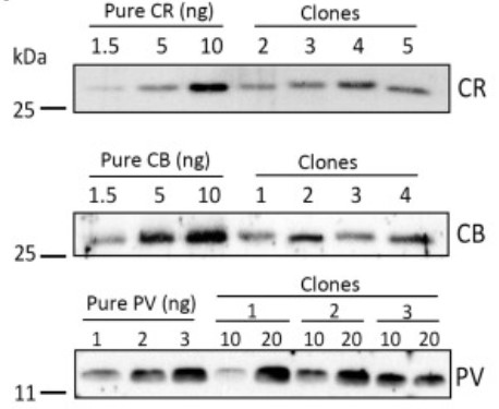
Fig1. Protein expression levels of CR, calbindin-D28k (CB), and parvalbumin (PV) in SPC111 clones obtained by serial dilution by Western blot analyses.
Case study 2: Valentina Medici, 2016
A consistent decrease in the number of inhibitory interneuronal subpopulation that expresses parvalbumin (PV) was reported in postsurgical tissue from patients with focal cortical dysplasia (FCD). The team tested if the decrease in PV protein expression observed in epileptic tissue corresponds to a parallel impairment in the γ-aminobutyric acid (GABA)ergic compartment.
Serial sections were processed using in situ hybridization with GAD-65 and GAD-67 probes and immunocytochemistry with antibody against PV. The density of inhibitory PV-immunoreactive interneurons in relation to GABAergic cells was estimated in controls. Field fraction and line profile analyses were added to estimate immunostaining proportion and distribution of PV signal generated in gray matter.
A reduction of PV-positive cells and PV-immunoreactivity was observed exclusively in FCD type I/III specimens compared with cryptogenic tissue from control patients with a poor postsurgical outcome. In FCD type II, a profound rearrangement in the cortical distribution of PV immunoreactivity was observed, without a quantitative reduction of the number of neurons and terminals.
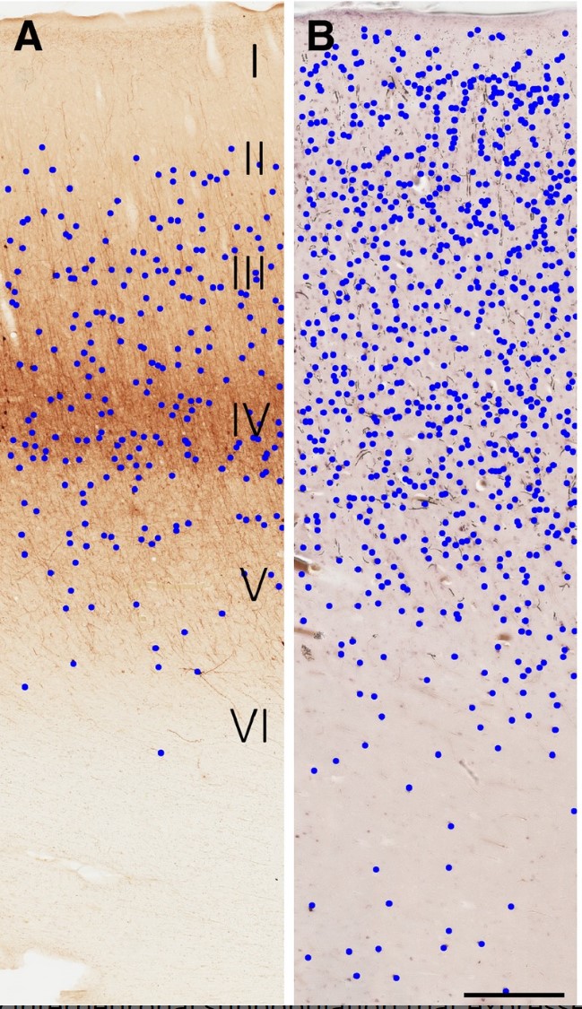
Fig3. Methodologic aspects of 2D (A–B) analyses of PV-positive and GAD65/67-positive neuronal density.
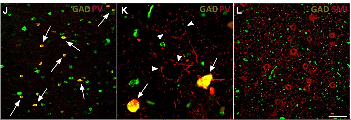
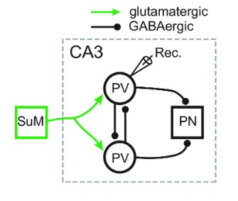
Fig2. The absence of monosynaptic GABAergic connection between SuM and CA3 PV+ neurons. (Minghua Li, 2023)
PVALB involved in several pathways and played different roles in them. We selected most pathways PVALB participated on our site, such as , which may be useful for your reference. Also, other proteins which involved in the same pathway with PVALB were listed below. Creative BioMart supplied nearly all the proteins listed, you can search them on our site.
| Pathway Name | Pathway Related Protein |
|---|
PVALB has several biochemical functions, for example, calcium ion binding, protein heterodimerization activity, protein homodimerization activity. Some of the functions are cooperated with other proteins, some of the functions could acted by PVALB itself. We selected most functions PVALB had, and list some proteins which have the same functions with PVALB. You can find most of the proteins on our site.
| Function | Related Protein |
|---|---|
| calcium ion binding | ICN;LCP1;NKD2A;GUCA1G;EPDL1;PCDHA4;PCDH2G28;PLS3;EFCAB6 |
| protein heterodimerization activity | PRPH;MYOD1;BAXB;JAM3;DR1;HIST1H4E;Pdgfa&Pdgfb;TTR;PEF1 |
| protein homodimerization activity | MAP3K12;SOD1;SLIT2;GRIK1;E2F8;VIL1;SERPINF2;SLC9A7;SRR |
PVALB has direct interactions with proteins and molecules. Those interactions were detected by several methods such as yeast two hybrid, co-IP, pull-down and so on. We selected proteins and molecules interacted with PVALB here. Most of them are supplied by our site. Hope this information will be useful for your research of PVALB.
- Q&As
- Reviews
Q&As (7)
Ask a questionPVALB (Parvalbumin) is a calcium-binding protein primarily expressed in fast-spiking inhibitory interneurons of the brain. Its main function is to regulate calcium signaling and buffering within neuronal cells. PVALB acts as a calcium buffer, preventing excessive calcium influx and maintaining proper calcium homeostasis. Additionally, PVALB is involved in modulating synaptic transmission, shaping neuronal network activity, and regulating neuronal excitability, ultimately contributing to the fine-tuning of neuronal circuits.
Emerging evidence suggests that alterations in PVALB expression and function may contribute to the pathogenesis of neurodevelopmental disorders, such as autism spectrum disorders (ASD) and schizophrenia. Reduced PVALB expression has been observed in postmortem brain tissues of individuals with ASD and schizophrenia. Animal models with PVALB deficiencies exhibit behavioral phenotypes resembling certain aspects of these disorders. Dysregulated inhibitory signaling mediated by PVALB interneurons can disrupt the excitatory-inhibitory balance in neural circuits, ultimately leading to cognitive and behavioral impairments associated with neurodevelopmental disorders.
Targeting PVALB has shown potential therapeutic implications in certain neurological disorders. Modulating PVALB expression or activity could potentially restore the disrupted excitatory-inhibitory balance observed in neurodevelopmental disorders. Strategies aimed at enhancing PVALB expression, such as epigenetic modifications or pharmacological interventions, could potentially alleviate cognitive and behavioral deficits in these disorders. However, further research is needed to fully understand the complexity of PVALB-mediated mechanisms in neurological disorders and to develop targeted therapies that specifically modulate PVALB function without interfering with normal neuronal function.
PVALB has been proposed as a potential biomarker for certain neurological diseases. Altered PVALB expression has been observed in several neurodegenerative disorders, including Alzheimer's disease and Parkinson's disease. However, the use of PVALB as a reliable biomarker requires further validation and standardization across different patient populations. Additionally, the development of sensitive and specific assays for measuring PVALB levels in biological samples is necessary to establish its clinical utility as a biomarker. Overall, while PVALB shows promise as a biomarker, more research is needed to establish its diagnostic and prognostic value in neurological diseases.
The expression of PVALB is regulated by various factors, including neuronal activity and transcriptional regulation. Increased neuronal activity, such as enhanced synaptic input, has been shown to upregulate PVALB expression. Transcription factors, such as Lhx6 and Nkx2.1, are involved in the regulation of PVALB gene expression. Additionally, epigenetic modifications, such as DNA methylation and histone acetylation, may also influence PVALB expression levels. However, further research is needed to fully elucidate the precise mechanisms underlying the regulation of PVALB expression in neuronal cells.
The expression of PVALB is tightly regulated during neuronal development and in response to specific physiological and pathological conditions. Studies have identified several transcription factors and signaling pathways involved in the regulation of PVALB expression. For instance, the activity-dependent transcription factor, cAMP response element binding protein (CREB), has been shown to bind to the promoter region of the PVALB gene and regulate its expression in a calcium-dependent manner. Additionally, the Wnt signaling pathway and various growth factors have also been implicated in the modulation of PVALB expression.
PVALB deficiency in the brain leads to various physiological consequences. It has been observed that the loss of PVALB function results in altered neuronal excitability and increased susceptibility to seizures. Impaired calcium buffering due to PVALB deficiency can disrupt synaptic plasticity, leading to deficits in learning and memory processes. Additionally, PVALB deficiency has been associated with imbalanced neuronal network activity and disruptions in the synchronization of neuronal firing patterns. These physiological consequences highlight the crucial role of PVALB in maintaining proper neuronal function and circuit dynamics.
Customer Reviews (3)
Write a reviewWith the assistance of this protein reagent, I am able to more accurately determine protein concentrations, resulting in enhanced precision in my experimental results.
This reagent is safe to use, without any safety concerns.
The manufacturer's proactive suggestions have revolutionized my experimental setup, leading to more accurate results.
Ask a Question for All PVALB Products
Required fields are marked with *
My Review for All PVALB Products
Required fields are marked with *