Skeletal System Development
Creative BioMart Skeletal System Development Product List
Immunology Background
Background
The development of the skeletal system is a complex process that involves the formation, growth, and maturation of bones in the human body. During embryonic development, the skeleton initially consists of cartilage and fibrous membranes. Over time, this cartilaginous skeleton is replaced by bone through a process called ossification.
Ossification is the process by which bone tissue is formed. There are two main types of ossification:
- Endochondral ossification: Replaces hyaline cartilage with bone, and is responsible for the formation of long bones during embryonic development. This process involves five steps, including the differentiation of mesenchymal cells into chondrocytes, which then secrete a matrix to form cartilage. Blood vessels then bring osteoblasts to the cartilage, and capillaries deposit bone inside the model. Finally, cartilage and chondrocytes continue to grow at the ends of the bone.
- Intramembranous ossification: Replaces connective tissue membranes with bone, and is responsible for the formation of flat bones in the skull and turtle shells. This process involves the proliferation of mesenchymal cells into compact nodules, and then osteoblasts migrating to the membranes and depositing bone matrix around themselves.
Markers of skeletal system development are specific molecules or substances that can be used to monitor bone formation, growth, and turnover. These markers provide valuable insights into bone health and can help in diagnosing and monitoring various bone disorders. Here are some key markers: alkaline phosphatase (ALP), osteocalcin, type I collagen, bone alkaline phosphatase (BAP), and osteopontin.
By monitoring these markers, clinicians and researchers can assess bone health, track bone development, diagnose bone-related disorders, and evaluate the effectiveness of treatments aimed at improving skeletal system function. Understanding the role of these markers in skeletal system development is crucial for maintaining and promoting bone health throughout life.

The Role and Regulatory Mechanisms of Skeletal System Development Markers
Skeletal system development markers play essential roles in monitoring and regulating the complex processes involved in bone formation, growth, and remodeling during skeletal development. These markers serve as indicators of various aspects of skeletal health and can help in diagnosing, assessing, and treating bone-related disorders. Here's a breakdown of the role and regulatory mechanisms of skeletal system development markers in skeletal development:
Role of Skeletal System Development Markers
| Role | Details |
|---|---|
| Monitoring Bone Formation and Turnover | Skeletal system development markers, such as osteocalcin, bone alkaline phosphatase (BAP), and type I collagen, serve as indicators of bone formation and turnover. They help track the activity of osteoblasts and osteoclasts, which are crucial for bone remodeling and maintenance. |
| Assessing Bone Health | These markers provide valuable information about bone health and density. Changes in their levels can indicate conditions like osteoporosis, osteomalacia, or Paget's disease, allowing for early detection and intervention. |
| Evaluating Bone Growth and Development | Skeletal system development markers can be used to monitor bone growth and development in children and adolescents. They help assess whether bone growth is occurring at a normal rate and identify any abnormalities or growth deficiencies. |
| Diagnosing Bone Disorders | Abnormal levels of skeletal system development markers can indicate various bone disorders or diseases. For example, elevated levels of tartrate-resistant acid phosphatase (TRAP) may suggest increased osteoclast activity associated with conditions like osteoporosis. |
Regulatory Mechanisms of Skeletal System Development Markers
| Mechanisms | Details |
|---|---|
| Gene Expression Regulation | The expression of genes encoding skeletal system development markers is tightly regulated by various transcription factors and signaling pathways. For example, transcription factors like Runx2 play a crucial role in regulating the expression of osteogenesis markers like osteocalcin. |
| Cell Signaling Pathways | Signaling pathways such as Wnt, BMP, and Notch signaling play key roles in regulating the activity of cells involved in skeletal development. These pathways can influence the expression and activity of skeletal system development markers. |
| Feedback Mechanisms | The levels of skeletal system development markers can influence the activity of bone-forming and bone-resorbing cells. For example, osteocalcin, a marker of bone formation, can regulate insulin secretion and energy metabolism, forming a feedback loop between bone and other physiological processes. |
| Environmental Factors | External factors such as diet, physical activity, hormonal balance, and exposure to certain substances can also influence the levels and activity of skeletal system development markers, affecting bone health and development. |
By understanding the roles and regulatory mechanisms of skeletal system development markers, researchers and healthcare providers can gain insights into bone development, health, and disease. Monitoring these markers can help in early diagnosis, treatment planning, and evaluation of therapies aimed at promoting skeletal health and function throughout life.
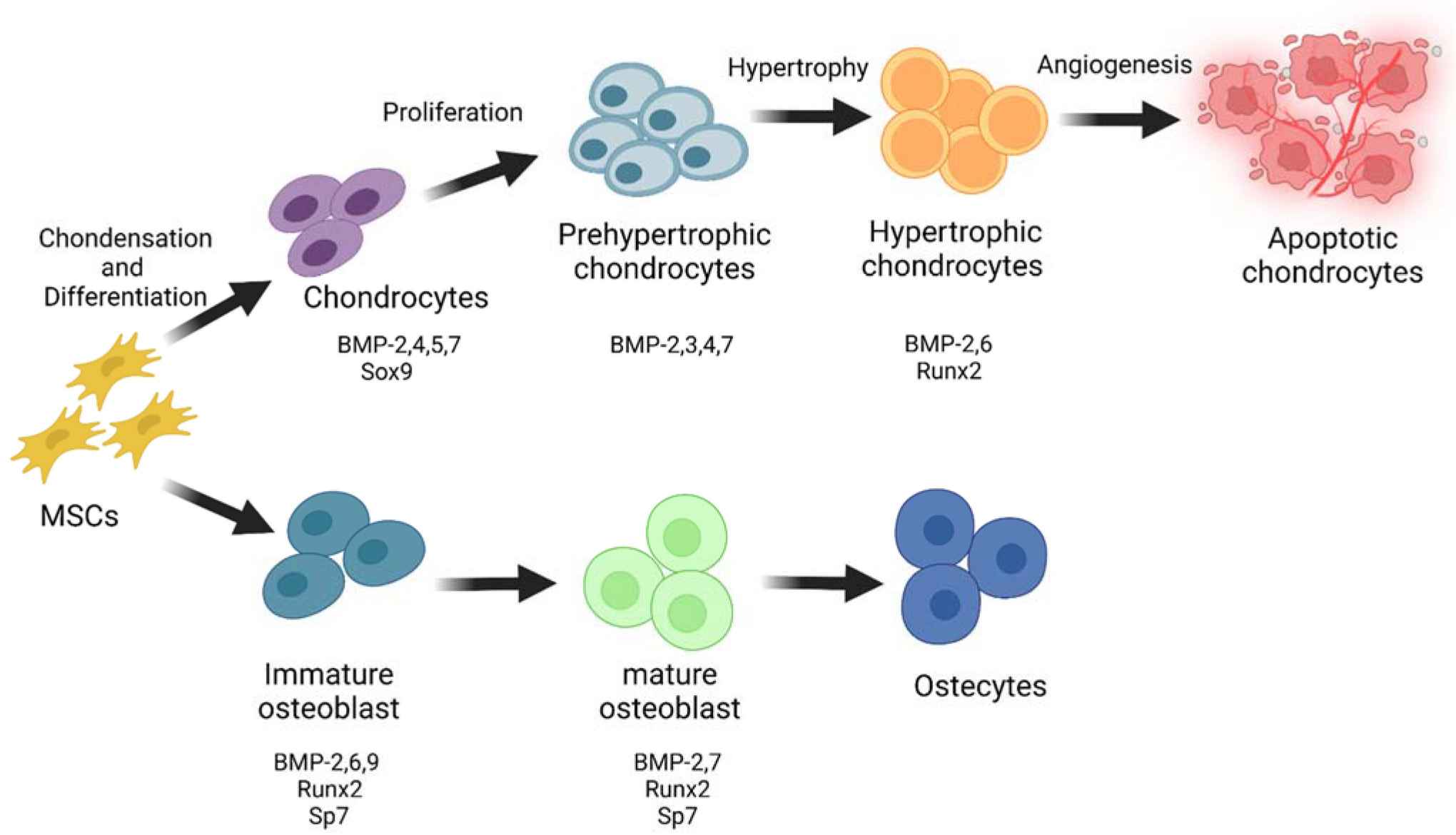
 Fig.1 Roles of BMPs in skeletal development. (Koosha E, et al., 2022)
Different Types of Skeletal System Development Markers
Markers play a crucial role in understanding the development and health of the skeletal system. Here are some key markers categorized based on different stages and processes of skeletal system development:
| Types | Features |
|---|---|
| Chondrogenesis Markers |
Chondrogenesis refers to the process of cartilage formation, which is essential for the development of the skeletal system, particularly during embryonic and fetal stages.
|
| Osteoclast Markers |
Osteoclasts are specialized bone cells responsible for bone resorption, a process crucial for bone remodeling and maintenance of bone density.
|
| Osteogenesis Markers |
Osteogenesis is the process of bone formation, which involves the differentiation of osteoblasts and the subsequent mineralization of the extracellular matrix to form bone tissue.
|
These markers provide valuable insights into the processes of chondrogenesis, osteoclast activity, and osteogenesis, helping researchers and healthcare providers monitor skeletal system development, bone health, and bone remodeling processes.
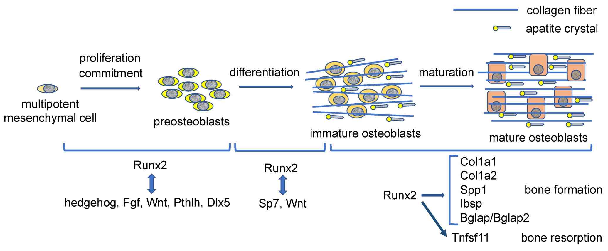
 Fig.2 Regulation of proliferation, differentiation, and bone matrix protein gene expression by Runx2 during osteoblast differentiation. (Komori T, 2022)
Clinical Applications of Skeletal System Development Markers
By utilizing skeletal system development markers in clinical practice, healthcare providers can enhance their diagnostic capabilities, tailor treatment approaches, and monitor bone health effectively. The levels of these markers in different diseases or conditions provide valuable insights into bone turnover, remodeling, and overall skeletal health, offering a holistic approach to health assessment that considers both bone and systemic health factors.
| Clinical applications | Details |
|---|---|
| Clinical Diagnosis |
|
| Treatment |
|
| Monitoring |
Cardiovascular Risk: Recent studies have linked markers like osteoprotegerin and osteocalcin to cardiovascular risk. Monitoring these markers alongside traditional cardiovascular risk factors can provide a more comprehensive health assessment. Metabolic Health: Markers such as sclerostin and fibroblast growth factor 23 (FGF23) are associated with metabolic disorders like diabetes and chronic kidney disease. Monitoring these markers can offer insights into both skeletal and metabolic health. |
Case Study
Case 1: Ichiyama-Kobayashi S, Hata K, Wakamori K, et al. Chromatin profiling identifies chondrocyte-specific Sox9 enhancers important for skeletal development. JCI Insight. 2024;9(11):e175486.
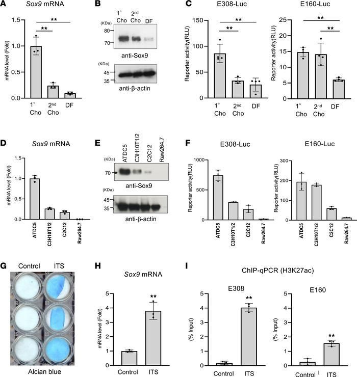
The transcription factor Sox9 is crucial for chondrogenesis and mutations in and around the gene can lead to campomelic dysplasia, a skeletal malformation disorder. Despite the well-established role of Sox9 in chondrocytes, the mechanisms controlling its expression have not been fully understood. Through genome-wide profiling, researchers have identified two enhancers, E308 and E160, located upstream of Sox9, which play a role in regulating its expression. Deleting both enhancers in mice resulted in a dwarf phenotype and reduced Sox9 expression in chondrocytes, affecting bone morphogenetic protein 2-dependent chondrocyte differentiation. Additionally, the loss of E308 and E160 led to a reorganization of an open chromatin region upstream of Sox9. These findings shed light on the regulation of the Sox9 gene in chondrocytes and may contribute to our understanding of skeletal disorders.
 Fig.1 Correlation of identified enhancer activity with Sox9 expression.
Related References
- Berendsen AD, Olsen BR. Bone development. Bone. 2015;80:14-18.
- Salhotra A, Shah HN, Levi B, Longaker MT. Mechanisms of bone development and repair. Nat Rev Mol Cell Biol. 2020;21(11):696-711.
- Komori T. Whole aspect of Runx2 functions in skeletal development. International Journal of Molecular Sciences. 2022; 23(10):5776.
- Koosha E, Eames BF. Two modulators of skeletal development: BMPs and proteoglycans. Journal of Developmental Biology. 2022; 10(2):15.

