


Intracellular Cytokine Detection and Activity Assay
Creative BioMart offers advanced Intracellular Cytokine Detection and Activity Assay services to provide highly sensitive and accurate measurement of cytokine production at the single-cell level. By combining intracellular cytokine staining with flow cytometry, we can identify functional antigen-specific T cells, characterize cytokine-secreting populations, and assess cellular responses with multiparameter analysis. This approach delivers deeper insights compared to conventional assays such as ELISA or immunohistochemistry, which cannot distinguish cytokine production at a per-cell resolution. Our customizable, cost-effective, and carefully optimized workflows are designed to accelerate immune research, drug development, and disease mechanism studies.
Understanding Intracellular Cytokine Detection and Immune Activity Assays
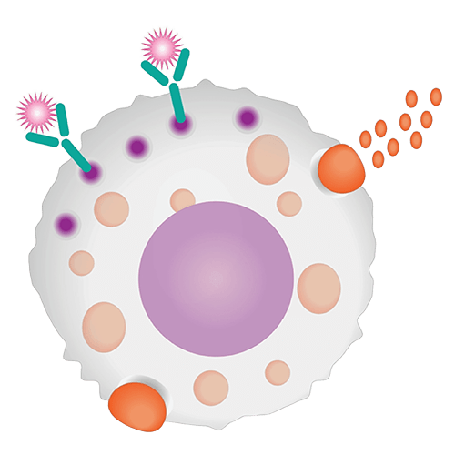
Cytokines are biologically active small proteins secreted by immune cells (e.g., monocytes, macrophages, T cells, B cells, NK cells) and non-immune cells (e.g., endothelial cells, fibroblasts, epidermal cells) in response to stimulation. These low molecular weight proteins regulate cell growth, differentiation, immune response, hematopoiesis, pluripotency, and tissue repair by binding specific receptors.
Cytokines include diverse families such as interleukins, interferons, tumor necrosis factors, colony-stimulating factors, chemokines, and growth factors. They play pivotal roles in pathways like IL-2 –induced T cell proliferation and TNF-α –mediated inhibition of viral gene expression. Dysregulation of cytokine expression is linked to numerous diseases, making their detection and quantification essential for both basic research and clinical applications.
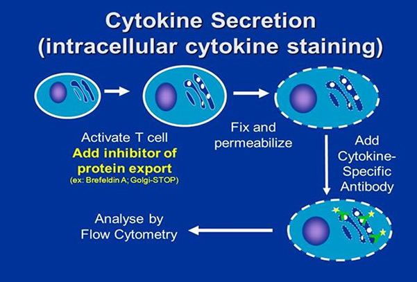
While traditional methods such as ELISA, limiting dilution analysis (LDA), and immunohistochemistry measure cytokines in bulk, they lack the resolution to analyze cytokine production at the single-cell level. Flow cytometry–based intracellular cytokine staining overcomes this limitation, enabling multiparametric analysis of cytokine-producing cell populations with unparalleled precision.
Comprehensive Cytokine Profiling and Functional Immune Assay Services
Creative BioMart provides a comprehensive range of services, including:
-

Single-Cell Cytokine Detection
Measure cytokine production at the individual cell level.
-

Functional T Cell Analysis
Detect antigen-specific T cell populations and assess functionality.
-

Multiparameter Flow Cytometry
Analyze up to 18 customizable parameters for detailed phenotyping.
-

Cytokine Secretion Confirmation
Characterize secretion profiles using antigen-specific cells.
-

Customizable Assay Design
Tailored strategies to fit project needs, balancing cost-effectiveness and scientific rigor.
-

Collaborative Support
Expert consultation for assay optimization and research-specific solutions.
Service Workflow

Service Details
|
Item |
Details |
|---|---|
|
Technology |
Flow cytometry with intracellular cytokine staining. |
|
Applications |
|
|
Customization |
Assay design, antibody panels, and experimental conditions tailored to client needs. |
|
Deliverables |
High-resolution multiparameter flow cytometry data with expert interpretation and reporting. |
Our Advantages in Cytokine Detection and Activity Assay
- Single-Cell Resolution: Detect cytokine-producing cells that bulk assays cannot reveal.
- High Multiplexing Capability: Up to 18 customizable markers analyzed in one experiment.
- Expert Assay Design: Tailored solutions to fit specific project needs.
- Cost-Effective Options: Optimized workflows that balance quality and affordability.
- Collaborative Approach: Ongoing consultation to refine and improve study outcomes.
- Reliable Results: Robust protocols ensuring reproducible and publication-ready data.
Research Applications and Case Studies in Cytokine Activity Analysis
Case 1: Intracellular cytokine detection in chronic inflammatory diseases
Zhang et al., 2023. doi:10.1111/all.15486
Understanding cytokine-driven immune responses at the single-cell level is vital for deciphering the mechanisms underlying chronic inflammatory diseases such as atopic dermatitis (AD) and psoriasis (PS). Using single-cell RNA sequencing and T-cell receptor (TCR) profiling, researchers identified disease-specific T-cell clusters that correlate with disease severity—Th2/Th22 in AD and Th17/Tc17 in PS. These inflammatory signatures extended beyond immune cells to fibroblasts, keratinocytes, and stromal cells in AD, while systemic type-3 inflammation was evident in PS. Additionally, macrophages showed antiviral activation linked to disease severity. This study highlights intracellular cytokine detection as a powerful tool for revealing disease-specific pathways and therapeutic targets.
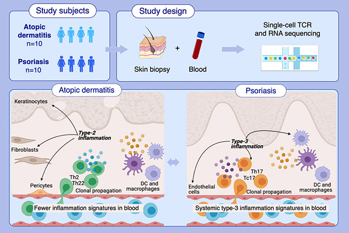
Figure 1. Single-cell RNA and TCR sequencing were applied to immune cells enriched from skin biopsies and matched blood samples of atopic dermatitis and psoriasis patients. The analysis uncovered disease-specific Th2/Th22 and Th17/Tc17 sub-populations in atopic dermatitis and psoriasis, respectively, with their abundance correlating with severity scores in both diseases. (Zhang et al., 2023)
Case 2: CD20+ T-cells in MGUS and multiple myeloma: dual roles in immunity
Forró et al., 2025. doi:10.3389/fimmu.2025.1464940
CD20+ T-cells, first described in blood and bone marrow, were investigated in patients with monoclonal gammopathy of undetermined significance (MGUS) and multiple myeloma (MM) compared with controls. Using flow cytometry and RNA sequencing, researchers found that CD20+ T-cells are phenotypically and transcriptionally distinct from CD20- T-cells. Their elevated incidence in MGUS and MM, along with expression of CD8, NKG2D, and CD28, points to antitumor functionality, while increased PD-1 expression indicates T-cell exhaustion in advanced disease. These cells are primarily effector or memory T-cells, suggesting a dual role in immune surveillance and tumor immune escape that influences MM progression.
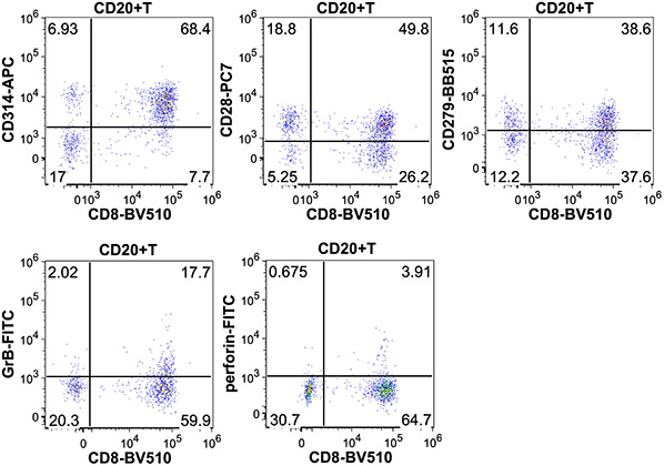
Figure 2. Representative dot plots showing the CD314 (NKG2D), CD28, CD279 (PD-1), granzyme-B and perforin expression of CD8+CD20+ T-cells. (Forró et al., 2025)
Client Experiences with Our Intracellular Cytokine Assay Services
“We collaborated with Creative BioMart to assess T cell functionality during the development of a novel influenza vaccine. Their intracellular cytokine detection assays provided clear identification of IL-2 and IFN-γ producing T cells at the single-cell level. The precision of their flow cytometry analysis confirmed robust immune activation, which became critical evidence in our preclinical dossier. The results gave our development team the confidence to move forward with regulatory submissions.”
— Director of Immunology Research | Global Vaccine Manufacturer
“Our team was investigating cytokine dysregulation in systemic lupus erythematosus, and Creative BioMart’s assay service proved invaluable. Their high-parameter flow cytometry revealed elevated IL-17 and TNF-α production in specific T cell subsets, providing mechanistic insights we could not achieve with ELISA alone. The data not only strengthened our publication but also shaped our therapeutic strategy moving forward.”
— Principal Investigator | Leading Academic Medical Center
“We partnered with Creative BioMart to evaluate cytokine profiles in patient-derived T cells undergoing checkpoint inhibitor treatment. Their intracellular cytokine staining workflow identified IFN-γ and IL-10 producing subsets, revealing the balance between immune activation and suppression. This level of detail was instrumental in refining our combination therapy approach. Their scientific team’s consultation throughout the project was as valuable as the data itself.”
— Senior Scientist, Translational Oncology | Mid-Sized Biotech Company
“During early-stage development of an immunomodulatory drug, we needed a cost-effective yet detailed assessment of cytokine responses. Creative BioMart designed a customized panel of 15 parameters, enabling us to track IL-4, IL-6, and IFN-γ simultaneously across multiple cell populations. The speed and reproducibility of their assays saved us months of work and gave us a reliable data package to support our IND application.”
— R&D Director | Pharmaceutical Company
FAQs About Cytokine Detection and Activity Assays
-
Q: What makes intracellular cytokine detection different from ELISA or other traditional methods?
A: Unlike ELISA or immunohistochemistry, which measure cytokines in bulk, intracellular cytokine detection allows analysis at the single-cell level. This provides detailed insights into which specific cell populations are producing cytokines, as well as their functional phenotypes. -
Q: Which cytokines can your assay detect?
A: Our assays can be customized to detect a wide panel of cytokines, including IL-2, IFN-γ, TNF-α, IL-4, IL-6, IL-10, IL-17, and others depending on the research needs. Multiplexing capabilities allow the analysis of up to 18 parameters simultaneously. -
Q: What types of cells can be analyzed with this assay?
A: We can analyze cytokine production from a variety of immune cells (T cells, B cells, NK cells, monocytes, macrophages) and antigen-specific cells, as well as cells derived from patient samples or in vitro systems. -
Q: How does the workflow ensure accurate cytokine measurement?
A: Our workflow includes stimulation of cells, inhibition of cytokine secretion, fixation, permeabilization, and intracellular staining with specific antibodies. Using high-parameter flow cytometry, we capture precise cytokine profiles and minimize artifacts caused by extracellular measurements. -
Q: Can the assay be tailored to my research project?
A: Yes. We design customized antibody panels, stimulation protocols, and gating strategies to match your study objectives, whether it’s vaccine development, autoimmune research, cancer immunotherapy, or drug screening. -
Q: How reliable are the results from your assays?
A: Our protocols are optimized for high reproducibility and sensitivity, and results are validated with internal controls. Clients receive detailed reports with expert interpretation to support publications, regulatory submissions, or further research. -
Q: What are the applications of intracellular cytokine detection?
A: This service is widely applied in:- Immune profiling for infectious diseases, autoimmune disorders, and cancer
- Evaluating T cell responses in vaccine development
- Characterizing cytokine dysregulation in disease pathways
- Screening and validating immunomodulatory drugs and biologics
Resources
Related Services
Related Products
References:
- Forró B, Kajtár B, Lacza Á, et al. Multiparameter flow cytometric and transcriptional analyis of CD20 positive T-cells in bone marrow in patients of multiple myeloma and monoclonal gammopathy of undetermined significance. Front Immunol. 2025;16:1464940. doi:10.3389/fimmu.2025.1464940
- Zhang B, Roesner LM, Traidl S, et al. Single‐cell profiles reveal distinctive immune response in atopic dermatitis in contrast to psoriasis. Allergy. 2023;78(2):439-453. doi:10.1111/all.15486
Contact us or send an email at for project quotations and more detailed information.
Quick Links
-

Papers’ PMID to Obtain Coupon
Submit Now -

Refer Friends & New Lab Start-up Promotions