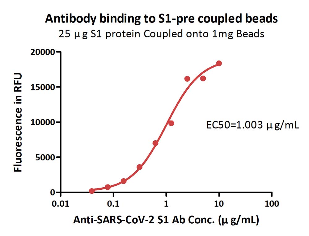
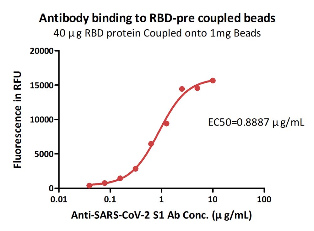
Pre-Coupling Magnetic Bead
🧪 HER2-006M
Source: HEK293
Species: Human
Tag:
Conjugation:
Protein Length: Thr 23 - Thr 652

🧪 EpCAM-016M
Source:
Species: Human
Tag:
Conjugation:
Protein Length:
Background
Overview
Pre-coupling magnetic beads are a widely used tool in bioscience research, especially in Immunoprecipitation (IP) experiments. These beads typically consist of A magnetic material (such as iron oxide) core and a chemically modified shell on which specific ligands, such as antibodies, protein A/G, biotin, or other affinity tags, can be coupled to achieve specific capture of the target molecule.

Features
High specificity: Ligands on pre-coupled magnetic beads can specifically recognize and bind to target proteins or nucleic acids, enabling highly selective separation.
Easy to operate: When experiments are conducted with pre-coupled magnetic beads, simple incubation and washing steps are usually required without complex sample pretreatment.
Fast and efficient: magnetic beads can be quickly separated by magnetic fields, greatly shortening the experiment time.
Low background: The design of the pre-coupled magnetic beads reduces the non-specific binding and improves the signal-to-noise ratio of the experiment.
Reagent saving: Due to the high affinity and specificity of magnetic beads, the use of reagents such as antibodies can be reduced, saving costs.
Automation compatibility: Pre-coupled magnetic beads are suitable for automation platforms that enable high-throughput screening and analysis.
Procedure of Use
Preparation of magnetic beads: Select the appropriate pre-coupled magnetic beads according to the experimental requirements, and pretreat them according to the instructions, such as washing and balancing.
Antibody binding: Antibodies are bound to magnetic beads, usually for a short incubation time.
Sample addition: The sample containing the target molecule is added to the magnetic bead and incubated to allow the target molecule to bind to the ligand on the magnetic bead.
Washing: Wash the magnetic beads with an appropriate buffer to remove unbound impurities.
Elution: The target molecule is eluted from the magnetic bead using an appropriate elution buffer as needed.
Follow-up analysis: SDS-PAGE, Western blot, mass spectrometry or other relevant analysis of the elution target molecules.
Development Course
The concept of pre-coupled magnetic beads was first proposed by chemist John Ugelstad at the Norwegian University of Science and Technology in 1976. He used polystyrene as the main material to create uniformly magnetized spherical particles, which laid the foundation for later magnetic bead technology. Subsequently, magnetic bead technology has been developed in the field of nucleic acid purification, especially in the 1990s, with the increase in the demand for high throughput, high sensitivity and automated operation in molecular biology and medicine, magnetic bead DNA extraction technology has been vigorously developed.
The precoupled magnetic beads combine the properties of immunology and superparamagnetic beads, which are superparamagnetic microspheres coated with antibodies or with antibody binding function. Such magnetic beads can rapidly gather under the action of an external magnetic field, and can be re-dispersed without remanence after the magnetic field is withdrawn. The ideal magnetic bead has a uniform spherical shape, composed of a superparamagnetic iron core and a protective polymer shell, with chemical functional groups on the surface, such as -OH, -NH2, -COOH and -CONO2, so that the magnetic bead can be coupled to almost any biologically active protein.
With the advancement of technology, the performance of pre-coupled magnetic beads is also constantly optimized. For example, Beckman Coulter's XP magnetic beads use SPRI (Solid Phase reversible immobilization) technology to improve the efficiency and purity of DNA adsorption. In addition, the surface modification and functionalization of magnetic beads are also constantly improved to adapt to different application needs.
The market for pre-coupled magnetic beads is growing with the development of biomedical research. With the emergence of new technologies, such as microfluidic and single-cell sequencing, the range of applications of pre-coupled magnetic beads is expected to expand further. In the future, pre-coupled magnetic bead technology may play a greater role in fields such as personalized medicine, precision diagnostics and biopharmaceuticals.
Applications
Immunoprecipitation (IP). Precoupled magnetic beads are widely used in immunoprecipitation experiments to specifically capture target proteins from complex biological samples. By pre-coupling antibodies to magnetic beads, target proteins can be quickly and efficiently extracted from cell lysates for subsequent protein analysis, such as Western Blot or mass spectrometry.
Chromatin Immunoprecipitation (ChIP). ChIP technology is used to study protein-DNA interactions, such as the identification of transcription factor binding sites. Pre-coupled magnetic beads can be used to capture DNA fragments that bind to specific proteins, such as histone modifying enzymes, thereby revealing the mechanisms of gene expression regulation.
Protein complex analysis. Precoupled magnetic beads can be used to capture specific protein complexes, helping researchers understand the interactions between proteins. This is essential to uncover the signaling pathways and molecular mechanisms of disease occurrence.
Nucleic acid purification. Precoupled magnetic beads can also be used for the purification of nucleic acids, for example, by coupling nucleic acid probes with specific sequences, it is possible to capture target DNA or RNA from complex samples. This is useful for applications such as gene expression analysis, mutation detection, and viral load monitoring.
Cell sorting. Pre-coupled magnetic beads can be used for cell sorting, and by coupling specific cell surface marker antibodies, specific types of cells can be isolated from mixed cell populations for cell biology research and therapeutic applications.
Biosensors. Pre-coupled magnetic beads can be integrated into biosensors to detect environmental contaminants, pathogens, or other biomarkers.
Clinical diagnosis. Pre-coupled magnetic beads also have important applications in clinical diagnosis, such as for pathogen detection, tumor marker analysis, and personalized medicine.
Case Study
Case Study 1: Streptavidin-coupled Magnetic Beads
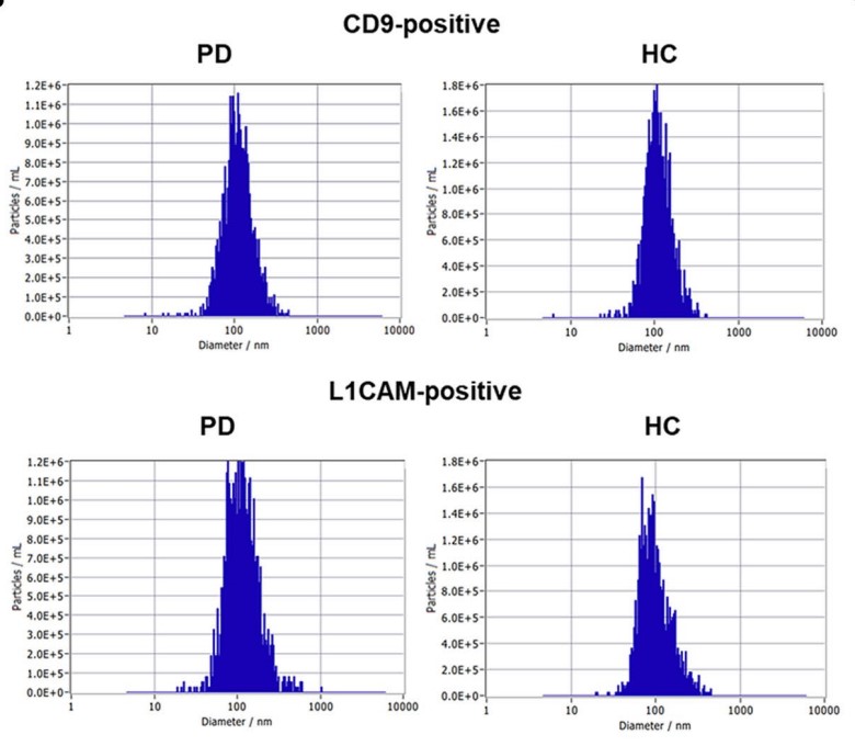
Parkinson's disease (PD) is a neurodegenerative disorder characterized by dopaminergic dysfunction and associated with abnormalities in the cholinergic system. However, the relationship between PD and cholinergic dysfunction, particularly in exosomes, is not fully understood. 37 patients with PD and 44 healthy controls (HC) were enrolled to investigate acetylcholinesterase (AChE) activity in CD9-positive and L1CAM-positive exosomes. Exosomes were isolated from plasma using antibody-coupled magnetic beads, and their sizes and concentrations were assessed using transmission electron microscopy, nanoparticle tracking analysis, and western blotting. A significant decrease in AChE activity was observed in CD9-positive exosomes derived from patients with PD, whereas no significant differences were found in L1CAM-positive exosomes. Further analysis with a larger sample size confirmed a substantial reduction in AChE activity in CD9-positive exosomes from the PD plasma, with moderate diagnostic accuracy.
(Sumin Jeong, 2024)
Fig1. Characteristics of the isolated CD9- or L1CAM-positive exosomes by magnetic beads conjugated with antibodies. The distribution of size and concentration of exosomes.
Case Study 2: GLAST antibody-coupled magnetic beads
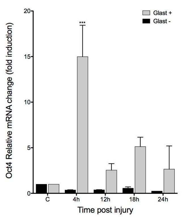
Müller glia (MG) is the most abundant glial type in the vertebrate retina. Among its many functions, it is capable of responding to injury by dedifferentiating, proliferating, and differentiating into every cell types lost to damage. This regenerative ability is notoriously absent in mammals. This might be explained by a mnemonic mechanism comprised by epigenetic traits, such as DNA methylation. To achieve a better understanding of this epigenetic memory, the expression of pluripotency-associated genes was studied, such as Oct4, Nanog, and Lin28, which have been reported as necessary for regeneration in fish, at early times after NMDA-induced retinal injury in a mouse experimental model. although Oct4 is expressed rapidly after damage (4 hpi), it is silenced at 24 hpi. By MS-PCR, a decrease was observed in Oct4 methylation levels at 4 and 12 hpi, before returning to a fully methylated state at 24 hpi. To demonstrate that these changes are restricted to MG, these cells were separated using a GLAST antibody coupled with magnetic beads.
(Luis I Reyes-Aguirre, 2016)
Fig2. qPCR quantification of Oct4 expression levels at the indicated time after injury in MACS GLAST-positive and negative fraction.
Case Study 3: Anti-GFP antibody-coupled magnetic beads
The majority of nuclear-encoded organellar proteins contain a cleavable presequence, which is necessary for protein targeting and import into the correct cellular compartment. Knowledge about targeting-peptide cleavage sites is essential for the structural and functional characterization of the mature organellar proteins as well as for a deeper understanding of the import process. Because of the low consensus and high variability of presequences, bioinformatics of targeting-peptide cleavage fails to predict the length of the targeting peptide with high confidence. Therefore, a rapid and robust method has been developed to experimentally determine the cleavage site of the transit peptide for proteins imported into mitochondria or plastids. The protein precursor with green fluorescent protein (GFP) fused to its C-terminus is transiently expressed in cells (for animal proteins) or protoplasts (for plant proteins), allowing translocation into organelles and removal of the transit peptide. After lysis, the matured protein is immunopurified using an anti-GFP antibody coupled to magnetic beads. The N-terminal amino sequence is then determined by Edman microsequencing or mass spectrometry.

(Adrien Candat, 2013)
Fig3. Western blot analysis of the immunopurified GFP-tagged proteins, using a monoclonal anti-GFP antibody.
Advantages
- Various products: We offer a wide variety of magnetic beads, including unmodified magnetic beads, universal specific ligand magnetic beads and specific recognition groups of magnetic beads, to meet the needs of different customers.
- Technical support: We will provide professional technical support and training to help customers optimize the experimental process and improve the efficiency of product use.
- Customized services: We are able to provide customized products and solutions according to the specific needs of our customers to enhance customer satisfaction and loyalty.
Creative BioMart contains a wide range of Pre-Coupling Magnetic Beads to provide an efficient, fast and economical method for the separation, detection and analysis of a wide range of biomolecules. You can also let us know if you have any customized requirements. Please contact us for more product details.
References
- Jeong S.; et al. Assessment of acetylcholinesterase activity in CD9-positive exosomes from patients with Parkinson's disease. Front Aging Neurosci. 2024;16:1332455.
- Reyes-Aguirre LI, Lamas M. Oct4 Methylation-Mediated Silencing As an Epigenetic Barrier Preventing Müller Glia Dedifferentiation in a Murine Model of Retinal Injury. Front Neurosci. 2016;10:523.
- Candat A.; et al. Experimental determination of organelle targeting-peptide cleavage sites using transient expression of green fluorescent protein translational fusions. Anal Biochem. 2013;434(1):44-51.


