


MAPK Signaling Pathway
🧪 KRAS-2569H
Source: E.coli
Species: Human
Tag: His
Conjugation:
Protein Length: 1-185 aa


🧪 EGF-04H
Source: E.coli
Species: Human
Tag:
Conjugation:
Protein Length:

🧪 IL1A-01H
Source: E.coli
Species: Human
Tag: Non
Conjugation:
Protein Length: 159
🧪 IL1B-02H
Source: E.coli
Species: Human
Tag: Non
Conjugation:
Protein Length: 153
🧪 IL1RN-05H
Source: E.coli
Species: Human
Tag: Non
Conjugation:
Protein Length: 152
🧪 EGFR-30H
Source: Human Cells
Species: Human
Tag: His
Conjugation:
Protein Length: 25-645 aa
🧪 CREB1-26H
Source: E.coli
Species: Human
Tag: Non
Conjugation:
Protein Length: 2-328 a.a.
🧪 CD40-493H
Source: HEK293
Species: Human
Tag: Fc
Conjugation:
Protein Length: 1-193 a.a.

🧪 CD27-638H
Source: Human Cells
Species: Human
Tag: Fc&His
Conjugation:
Protein Length: 1-192 a.a.

🧪 CD40-174H
Source: Human Cells
Species: Human
Tag: Fc&His
Conjugation:
Protein Length: 1-193 a.a.

🧪 FGFR1-138H
Source: HEK293
Species: Human
Tag: Fc&His
Conjugation:
Protein Length: 1-285 aa

🧪 EGFR-692H
Source: HEK293
Species: Human
Tag: Fc
Conjugation:
Protein Length: 1-640 aa

🧪 FGFR2-705H
Source: HEK293
Species: Human
Tag: Fc&His
Conjugation:
Protein Length: 1-377 aa

🧪 FGFR4-708H
Source: HEK293
Species: Human
Tag: Fc
Conjugation:
Protein Length: 22-369 aa

🧪 Fgfr4-709M
Source: HEK293
Species: Mouse
Tag: His
Conjugation:
Protein Length: Leu17-Asp366
Background of MAPK Signaling Pathway
What is MAPK Signaling Pathway?
Mitogen-activated protein kinase (MAPK) is a highly conserved family of serine/threonine protein kinases that play a role in many fundamental cellular processes, such as proliferation, differentiation, motility, stress response, apoptosis, and survival. Conventional MAPKs include extracellular signal-regulated kinases 1 and 2 (Erk1/2 or p44/42), c-Jun amino-terminal kinase 1-3 (JNK1-3)/ stress-activated protein kinase (SAPK1A, 1B, 1C), p38, and Erk5.
A wide range of extracellular stimuli, including mitogen, cytokine, growth factor, and environmental functional stimulators, stimulate the activation of one or more MAPKK kinases (MAPKKK) through receptor-dependent and receptor-independent mechanisms. After that, MAPKKK phosphorylates and activates the downstream MAPK kinase (MAPKK), which in turn phosphorylates and activates MAPK. Activation of MAPK leads to phosphorylation and activation of a specific MAPK-activated protein kinase (MAPKAPK).
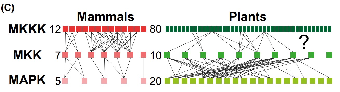
Fig1. Schematic representation of MAPK cascade in mammals and plants. (Yan Ma, 2023)
Core Components and Functions
MAPKKK acts as the starting point for the MAPK signaling pathway, receiving signals from outside the cell, such as growth factors, cytokines, or stress signals, and initiating signaling via proteins associated with membrane receptors, such as G proteins or receptor tyrosine kinases. There are several known MapKKKs, which belong to different subfamilies; for example, the Raf family (including B-RAF, A-RAF, and RAF-1) is A typical MAPKKK in the ERK pathway. MAPKKK activates MAPKK (MEK or MKK) by phosphorylating specific threonine and/or serine residues, a process that is a key step in the MAPK signaling pathway.
Located upstream of the MAPK signaling pathway, MAPKK receives activation signals from MAPKK kinase (MAPKKK or MKKK) and passes this signal to the downstream MAPK. MAPKK has typical protein kinase domains, and their activation requires phosphorylation of MAPKKK. There are many types of MAPKKs that can activate different MAPK subfamilies. For example, MEK1 and MEK2 are upstream kinases of ERK, while MKK3 and MKK6 are upstream kinases of p38 MAPK. MAPKK activates MAPK by diphosphorylation of specific threonine and tyrosine residues on its activation ring. This diphosphorylation is the result of phosphorylation of MAPK by MAPKK.
The MAPK family is divided into four main subfamilies based on their structure and function: ERK (extracellular signal-regulated kinase), p38 MAPK, JNK (c-Jun amino-terminal kinase), and ERK5 (extracellular signal-regulated kinase 5). MAPK activation is usually triggered by extracellular signals such as growth factors, cytokines, or stress signals. These signaling molecules bind to receptors on the cell surface and activate MAPKKK downstream, which in turn activates MAPKK and MAPK. Among them, ERK pathway is mainly related to cell proliferation and differentiation. JNK and p38 MAPK pathways play a role in cellular stress responses such as inflammation and apoptosis. The ERK5 pathway is involved in cell proliferation and survival.
Roles of MAPK Signaling Pathway
Cell Proliferation and Differentiation: The ERK (Extracellular signal-regulated kinase) branch of the MAPK pathway is particularly well-known for its role in promoting cell growth and differentiation. Activation of ERK can lead to the expression of genes involved in cell cycle progression and cell growth, making it a key player in development and tissue homeostasis.
Stress Response: The JNK (c-Jun N-terminal kinase) and p38 MAPK pathways are activated in response to various forms of cellular stress, including UV radiation, heat shock, and osmotic stress. They are involved in the regulation of genes that help the cell to respond and adapt to these stressors.
Inflammation: Both JNK and p38 MAPK are integral to the inflammatory response. They can activate transcription factors such as AP-1, which in turn regulate the expression of inflammatory cytokines and chemokines.
Apoptosis: The MAPK pathway also plays a role in programmed cell death or apoptosis. Activation of JNK and p38 MAPK can lead to the activation of pro-apoptotic proteins, contributing to the initiation of cell death pathways.
Cell Migration and Invasion: The ERK pathway is involved in cell migration and invasion, processes that are particularly significant during embryonic development and wound healing, but also in the metastatic spread of cancer cells.
Cellular Senescence: The MAPK pathway has been implicated in the regulation of cellular senescence, a state of irreversible cell cycle arrest that occurs in response to various stressors and can contribute to aging and age-related diseases.
Pathway Related Diseases
Cancer: Abnormal activation of the MAPK pathway is frequently observed in cancer, and mutations in components of the pathway, such as B-RAF, are associated with the development of certain types of cancer. The pathway is a target for cancer therapeutics due to its role in tumor cell survival and proliferation.
Neurodegenerative Diseases: The JNK pathway has been linked to neurodegenerative diseases such as Alzheimer's and Parkinson's disease, where it may contribute to neuronal cell death.
Cardiovascular Disease: p38 MAPK is involved in the regulation of cardiovascular function and has been implicated in the pathology of conditions such as heart failure and atherosclerosis.
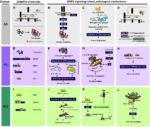
Fig2. Roles of MAPK pathways in the pathogenesis of Alzheimer's disease (AD), Parkinson's disease (PD), and amyotrophic lateral sclerosis (ALS). (Eun Kyung Kim, 2010)
Case Study
Case Study 1: Recombinant Human TNF protein (TNF-412H)
This study aims to evaluate the effects that early and late systemic administration of methylprednisolone have on lungs in a rat model of brain death. Twenty-four male Wistar rats were anesthetized and randomly divided into four groups: sham-operated (sham); brain death only (BD); brain death plus methylprednisolone (30 mg/kg i.v.) after 5 min (MP5); and brain death plus methylprednisolone (30 mg/kg i.v.) after 60 min (MP60). The researchers determined hemodynamic and arterial blood gas variables; wet/dry weight ratio; histological score; levels of thiobarbituric acid reactive substances (TBARS); superoxide dismutase (SOD) activity; and catalase activity. In BAL fluid, we determined differential white cell counts, total protein, and lactate dehydrogenase levels. Myeloperoxidase activity, lipid peroxidation, and TNF-α levels were assessed in lung tissue. The results showed that the levels of TBARS were significantly higher in the MP5 and MP60 groups than in the sham and BD groups (p < 0.001). The levels of TNF-α were significantly lower in the MP5 and MP60 groups than in the BD group (p < 0.001).
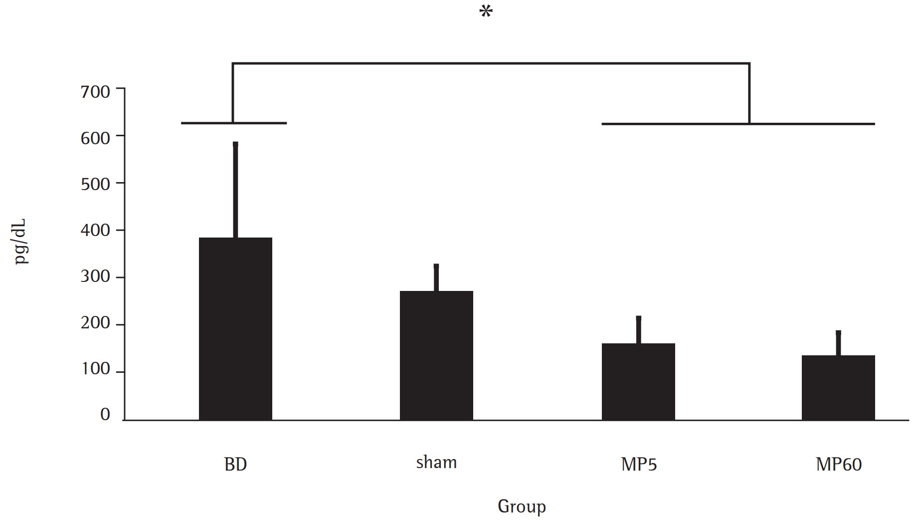
Fig1. TNF-α levels in lung tissue. (Eduardo Sperb Pilla, 2013)
Case Study 2: Recombinant Human TNFRSF11B protein (TNFRSF11B-487H)
The researchers explored the effects of different polymer structures on protein activity, specifically targeting Osteoprotegerin (OPG) activity. OPG is a protein that inhibits bone absorption by preventing the formation of mature osteoclasts. Due to increased osteoclast production, accelerated bone loss diseases such as osteoporosis, rheumatoid arthritis, and metastatic bone disease can lead to severe weakening of bones. Although OPG has shown promise in the treatment of bone diseases, its rapid liver clearance in the body limits its effectiveness and requires frequent and high dose administration to achieve therapeutic results.
In order to improve the efficacy of OPG, polymers with different branch Chain densities were prepared by Reversible Addition-Fragmentation Chain Transfer Polymerization (RAFT). A linear, loosely branched to densely branched polymer is included and conjugated with OPG to form an OPG-polymer bioconjugate. These bioconjugates were characterized by sodium dodecyl sulfate polyacrylamide gel electrophoresis (SDS-PAGE). In vitro experiments showed that OPG-polymer bioconjugates remained active against osteoclasts, and all conjugates were non-toxic. Preliminary in vivo studies further confirmed the non-toxic properties of these bioconjugates, with bone mineral density in rats measured by peripheral quantitative computed tomography (pQCT) at 7 days post-administration indicating a slight increase in bone mineral density after treatment with loosely branched OPG-polymer bioconjugates.
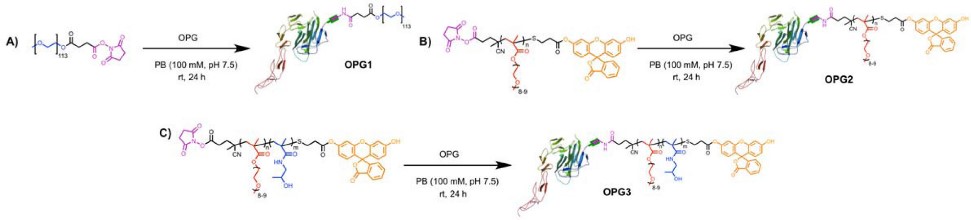
Fig2. OPG Bioconjugations with (A) P1 To Form OPG1, (B) P2 To Form OPG2, and (C) P3 To Form OPG3. (Bryan S Tucker, 2015)
Case Study 3: Recombinant Human HSP90B1 protein (HSP90B1-132H)
This study revealed the inhibitory effect of HCP1, a novel HSP90 inhibitor, on the apoptosis of vascular endothelial cells, providing a new strategy for the treatment of cardiovascular diseases. It was found that low dose of HCP1 can effectively reduce VEC apoptosis caused by lack of serum and FGF-2. HCP1 exerts anti-apoptotic effect by directly binding to Grp94 in endoplasmic reticulum and specifically inhibiting its activity. In addition, the overexpression of Grp94 can counteract the anti-apoptotic effect of HCP1, suggesting that Grp94 plays an important role in VEC apoptosis. This study not only provides insight into the molecular mechanism of VEC apoptosis, but also lays the foundation for the development of novel therapeutic agents targeting VEC apoptosis.
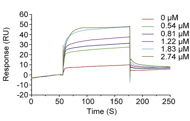
Fig3. Interaction of HCP1 with Grp94 was measured by using SPR analysis. (Qun Wei, 2019)
Related Products
MAPK signaling pathways are cascades of three kinases, where the most upstream kinase (MAPKKK) responds to various extra- and intracellular signals and activates the middle kinase (MAPKK) by direct phosphorylation. MAPKKs exclusively phosphorylate and activate a MAPK, which typically has many substrates that execute specific cell fate decisions adequate to the input signal. Focusing on the life sciences sector, Creative BioMart offers a range of high-quality products related to the MAPK signaling pathway. Please contact us if you have any need.