


Fluorescent Tagged Protein in Vivo
Creative BioMart provides comprehensive Fluorescent Tagged Protein In Vivo solutions built on our advanced ColorProbe™ fluorescent protein platform. Engineered for exceptional brightness, rapid maturation, and robust expression, ColorProbe™ enables precise visualization of protein localization, translocation, interaction, and gene expression in living systems. With a full spectrum of colors from blue to far-red, the platform supports multicolor labeling, co-localization analysis, and FRET-based interaction studies. Our service includes vector customization, fusion construction, and expression support in both bacterial and mammalian systems, ensuring reliable performance for in vivo imaging and functional studies.
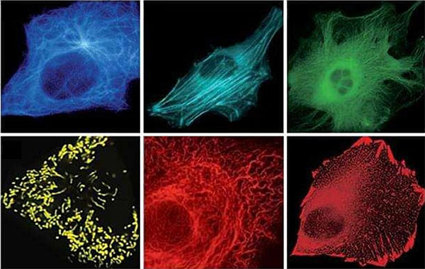
Introduction to ColorProbe™
Fluorescent protein tagging has become a cornerstone technique for studying protein behavior in living cells and organisms. By genetically fusing a fluorescent tag to the protein of interest, researchers can observe real-time dynamics such as trafficking, oligomerization, signaling, and organelle association. Creative BioMart’s ColorProbe™ fluorescent protein series is engineered to overcome common limitations of traditional fluorescent proteins—such as slow folding, low brightness, and poor performance in complex cellular environments.
Our long-standing track record demonstrates the reliability of ColorProbe™ in diverse model systems, including those involving highly oligomerizing and structurally sensitive cellular proteins. These optimized constructs allow researchers to perform multicolor imaging, FRET interaction assays, promoter activity monitoring, and whole-body imaging with exceptional clarity and consistency.
What Our ColorProbe™ Fluorescent Protein Platform Deliver
Services & Capacities
Creative BioMart provides a complete suite of in vivo fluorescent tagging services, including:
- ColorProbe™ Fluorescent Protein Collection spanning blue to far-red wavelengths
- Custom Fluorescent Protein Fusion Construction
- Vector Engineering and Optimization:
- Bacterial expression vectors
- Mammalian expression vectors
- Promoterless vectors
- Mitochondria-targeting vectors
- Ready-to-use subcellular localization vectors
- Stable Cell Line Expression Support
- In Vivo Imaging-Ready Labels suitable for multicolor studies, co-localization, and FRET assays
- Expert consultation on construct design and experimental strategy
Our Fluorescent Labeling In Vitro Service includes a complete suite of experimental steps and analytical support:
Core Labeling Services
- Conjugation of 1–5 mg of protein or antibody
- Universal C- or N-terminal labeling
- Dual-color labeling for advanced imaging or multiplex detection
- Amino- or thiol-reactive labeling depending on protein chemistry
Service Workflow

Service Features
-
ColorProbe™ Fluorescent Proteins
- Expanded color range for multiplex imaging in vivo
- Suitable for:
- Protein localization mapping
- Co-localization studies
- FRET-based interaction analysis
- Monitoring promoter activation
- Whole-body imaging in live organisms
-
Genetic Construct Options
- Single construct multicolor capability, enabling sophisticated live-cell experiments
- Subcellular targeting vectors for mitochondria and other organelles
- Promoterless constructs for custom regulatory element insertion
- Vector packages compatible with a wide range of hosts
-
Performance Attributes
- Superbright, fast-maturing fluorescence
- High stability across varied physiological environments
- Robust expression even in challenging systems
- Proven compatibility with highly oligomerizing proteins
Why Choose Our Platform
- Extensive ColorProbe™ Portfolio: Broad spectral options optimized for in vivo dynamics, multicolor assays, and FRET applications.
- Proven Functional Reliability: Years of application success in diverse cellular and organismal models, including challenging target proteins.
- Customizable Vector Engineering: Comprehensive solutions covering bacterial, mammalian, promoterless, and organelle-targeting constructs.
- High-Performance Fluorescence: Fast maturation, high brightness, and excellent photostability for reliable long-term imaging.
- Integrated Expression Support: Optional generation of stable cell lines ensures consistent and reproducible experimental outcomes.
- Expert Guidance from Start to Finish: Dedicated technical specialists help optimize design choices, imaging strategies, and downstream workflows.
Real-World Examples of Fluorescent Tagged Protein In Vivo
Case 1: Visualizing of the cellular uptake and intracellular trafficking of exosomes by live-cell microscopy
Tian et al., 2010. doi:10.1002/jcb.22733
Exosomes play vital roles in intercellular communication and show growing promise in diagnostics and therapeutics, yet their cellular uptake and trafficking remain incompletely understood. In this study, exosomes from PC12 cells were fluorescently labeled and incubated with resting PC12 cells to visualize their behavior. Live-cell microscopy revealed that exosomes enter cells via endocytosis, become enclosed in vesicles, and migrate toward the perinuclear region, likely through cytoskeleton-mediated active transport. Membrane proteins from exosomes were subsequently released and routed to lysosomes, while lipophilic dyes displayed reverse movement toward the cell periphery, suggesting lipid recycling. These findings provide new insight into exosome internalization and intracellular dynamics.
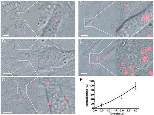
Figure 1. A–E: Time-lapse images of PC12 cells incubated with DiD-labeled exosomes (red) within 3 h. Merge of bright field with wide-field fluorescence images at 5 min (A), 30 min (B), 1 h (C), 2 h (D), and 3 h (E) after exosomes adding, respectively. Scale bar, 15 mm. F: Curve of exosome uptake dynamics by determining the fluorescent intensity. (Tian et al., 2010)
Case 2: Sulphide activity-dependent multicolor emission dye and its applications in in vivo imaging
Zhang et al., 2021. doi:10.1039/D1AN01345A
Reactive sulfur species (RSS) are essential in numerous biological processes, yet their interconnected reactivity patterns remain insufficiently characterized. To address this, researchers developed a multi-site fluorescent probe combining benzene-conjugated benzopyrylium with NBD, enabling systematic comparison of RSS behavior. The probe revealed distinct reaction pathways: SO2 cleaves the ether bond and additionally adds to the benzopyrylium double bond; GSH shows weaker dual reactivity, while highly reactive Cys/Hcy can further rearrange with NBD. These reactions generate multicolor emissions, enabling discrimination among sulfide species. The probe successfully visualized mitochondrial sulfides and supported cellular and zebrafish imaging, offering strong potential for practical diagnostic and analytical applications.
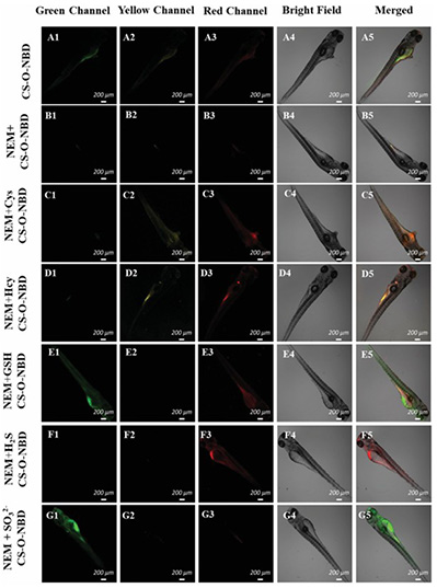
Figure 2. Fluorescence images of CS-O-NBD in zebrafish. (A) Zebrafish were incubated with CS-O-NBD (10 μM, 30 min). (B) NEM-pretreated (1 mM, 30 min) zebrafish were incubated with CS-O-NBD (10 μM, 30 min). (C–G) NEM-pretreated (1 mM, 30 min) zebrafish were incubated with Cys/Hcy/ GSH/H2S/SO32- (500 μM, 30 min), respectively, and then further incubated with CS-O-NBD (10 μM, 30 min). (Zhang et al., 2021)
What Our Clients Say
“We worked with Creative BioMart to develop a ColorProbe™-tagged fusion construct for monitoring the nuclear translocation of a transcription factor implicated in autoimmune disease. Their team engineered a mammalian vector that preserved the protein’s native regulation while delivering exceptionally bright and fast-maturing fluorescence. The constructs performed flawlessly during live imaging and enabled us to quantify translocation kinetics with unprecedented clarity.”
— Director of Immunology Research | Global Biotherapeutics Company
“Our oncology division needed a far-red fluorescent reporter system for whole-body imaging in a mouse xenograft model. Creative BioMart designed a customized ColorProbe™ fusion and provided a stable cell line expressing the construct at uniform levels. The brightness and photostability of the signal significantly improved our tumor progression tracking and reduced the number of imaging repeats required. Their work accelerated our in vivo screening timeline by several weeks.”
— Head of Translational Oncology | International Pharmaceutical Enterprise
“We collaborated with Creative BioMart on a project mapping mitochondrial dynamics in cardiomyocytes. Their team supplied a mitochondria-targeting fluorescent vector that integrated perfectly into our CRISPR workflow. The fluorescence intensity remained robust under high-stress culture conditions, and the rapid maturation allowed us to follow real-time fusion–fission events with high temporal resolution.”
— Principal Investigator, Cardiovascular Biology | Academic Medical Center
“For a developmental biology study, we needed multiple promoterless fluorescent constructs to dissect stage-specific gene expression in zebrafish embryos. Creative BioMart provided custom-designed ColorProbe™ reporters covering three different spectral channels. The resulting lines showed stable, clean expression without ectopic background, making our imaging quantification extremely reliable. Their technical support during vector design was exceptional.”
— Senior Scientist, Developmental Genetics | National Research Institute
Common Questions About Fluorescent Tagged Protein In Vivo
-
Q: What makes ColorProbe™ fluorescent proteins superior to traditional fluorescent tags?
A: ColorProbe™ proteins are engineered for exceptional brightness, fast maturation, and stability in a wide range of cellular environments. Unlike many conventional fluorescent proteins, they perform reliably in vivo —even in challenging systems involving highly oligomerizing or structurally sensitive proteins. Their broad spectral coverage also enables multicolor labeling, co-localization mapping, and FRET applications with high clarity. -
Q: Can you help me choose the best fluorescent protein for my specific in vivo application?
A: Absolutely. Our experts evaluate each project’s imaging requirements, expression system, spectral preferences, and protein characteristics to recommend the ideal ColorProbe™ variant. Whether you're conducting subcellular localization, whole-body imaging, promoter activity tracking, or protein interaction studies, we provide tailored guidance to ensure optimal signal performance. -
Q: Do you offer customizable vectors for different expression systems?
A: Yes. We provide an extensive range of expression vectors, including bacterial and mammalian vectors, promoterless constructs for regulatory studies, mitochondria-targeting vectors, and ready-to-use subcellular localization vectors. All vectors can be fully customized to meet your project requirements, including specific promoters, fusion orientations, or targeting sequences. -
Q: Will the fluorescent tag affect the biological function of my protein?
A: We use optimized fusion strategies and validated ColorProbe™ constructs designed to preserve the native function, localization, and regulation of your protein of interest. Our long track record includes successful tagging of proteins that are highly sensitive, oligomerization-prone, or involved in complex signaling pathways. Functional integrity is assessed as part of our quality control. -
Q: Can you generate stable cell lines expressing the fluorescent fusion protein?
A: Yes. For clients who require long-term, consistent expression, we offer complete stable cell line development services. Our team ensures uniform expression levels, robust fluorescence, and suitability for live imaging, high-content screening, or in vivo xenograft studies. -
Q: What types of in vivo studies are your ColorProbe™ tags compatible with?
A: ColorProbe™ fluorescent tags support a wide range of applications, including:- Real-time protein localization and trafficking
- Protein–protein co-localization
- FRET-based interaction measurements
- Visualization of promoter activation patterns
- Organelle-specific imaging
- Whole-body imaging in small animals
- Separation of mixed cell populations
-
Q: How do you ensure high brightness and signal consistency in vivo ?
A: ColorProbe™ proteins are engineered for rapid folding and strong fluorescence under physiological conditions. Through optimized vector construction, expression testing, and QC workflows, we ensure that the final constructs deliver high signal-to-noise ratios, minimal background expression, and reliable performance across experiments.
Other Resources
Related Services
- Protein Engineering Services
- Protein Interaction Service
- Protein Expression and Purification Services
- Bacterial Expression Systems ( E. coli / Bacillus)
- Mammalian Expression Systems
Related Products
References:
- Tian T, Wang Y, Wang H, Zhu Z, Xiao Z. V isualizing of the cellular uptake and intracellular trafficking of exosomes by live ‐cell microscopy. J of Cellular Biochemistry. 2010;111(2):488-496. doi:10.1002/jcb.22733
- Zhang Y, Wen L, Zhang W, et al. Sulphide activity-dependent multicolor emission dye and its applications in in vivo imaging. Analyst. 2021;146(18):5517-5527. doi:10.1039/D1AN01345A
Contact us or send an email at for project quotations and more detailed information.
Quick Links
-

Papers’ PMID to Obtain Coupon
Submit Now -

Refer Friends & New Lab Start-up Promotions