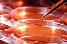
Cell culture
Cell culture introduction
 Most of animal cell lines grow on substrate as monolayer only a single thickness. In this case, once the all the surface of substrate is covered by cells, the cells stop growing and then following by differentiation or death. So it’s necessary to subculture cells regularly. In order to break the interactions between cells, trypsin or other enzymes are used. After the digestion of enzyme, cells will distribute in the medium homogeneously. Thus, a sort of cells can be transferred to new containers and begin to grow again until all the surface of substrate is covered by cells. The most important thing for cell culture is forbidden contamination from bacteria, fungi and mycoplasma and cross contamination with other cell lines. Anyway, please wear gloves, gauze mask and protective suit to make sure of your safety. This following protocol contains basic steps for cells culture.
Cell culture protocol
Materials
Marking pen
Waste container
CO2 incubator
Primary culture or subculture
Sterile PBS (pH 7.4)
Trypsin (pre-heat to 37 ºC)
Complete medium (pre-heat to 37 ºC): DMEM with 10% FBS (usually antibiotics added)
Pipettors
Incubator for 37 ºC warming
Cell counter
Cell culture plates /dishes
Sanitize
- Sanitize the inner surface of benchtop with 70% ethanol before work.
- Sanitize gloves with 70% ethanol and air dry for 30 seconds before work.
- Sanitize the exterior surfaces of all materials and equipment with 70% ethanol and transfer them into the benchtop before work.
- Sanitize the benchtop with 15– 30 min UV light.
- During the experiment, do not touch anything outside the benchtop (especially your skin and hair). Once gloves become contaminated, re-sanitize with 70% ethanol as above.
- Please move slowly within and outside the benchtop to allow the air within the benchtop to circulate properly.
- When finished, remove all the things outside the benchtop and sanitize the inner surfaces of benchtop with 70% ethanol.
- Sanitize the benchtop with 15– 30 min UV light.
Resuscitation of Frozen Cell Lines
- Remove cell in the container from liquid nitrogen and immediately place in a waterbath at 37ºC.
- As usual, it takes1-2 minutes to thaw the ice.
- Centrifuge the cells at 500 rpm for 2 min and transfer cells to the benchtop.
- Discard the supernatant and resuspend cells in1 mL medium gently.
- Transfer cells into culture dish and incubate for 24 h.
- Observe cells microscopically as necessary.
Subculture of Adherent (monolayer) Cell Lines
- Evaluate the degree of confluency of cell cultures with a microscope and make sure that cells grow well without bacterial and fungal contaminants.
- Remove the medium.
- Wash the cell with 3 ml of PBS (cells are culture in 10 cm´cm dishes).
- Dropwise add 1ml trypsin onto the washed cell monolayer using pipettor. Rotate dish to make all cells contact with trypsin.
- Incubate for 1-5 min according to the cell type until almost every cell becomes spherical and start to sheet.
- Add 2 mL medium with 10% FBS directly to inactivate the trypsin and resuspend the cells.
- Evaluate the cell concentration by cell counting.
- Add required number of cells (usually 0.6-0.8 ml) to a new labeled dish with 10 mL medium (total volume).
- Culture cell in a 37 ºC, humidified CO2 incubator.
- Observe cells microscopically and change medium as necessary

