


Principle and Protocol of BCA Method
BCA method is one of the most commonly used protein concentration detection methods in the world at present. It is not affected by ionic and non-ionic detergents in the sample, and the coefficient of variation for detecting different protein molecules is far less than Coomassie brilliant blue method. The determination can be completed within 45 min, which is 4 times faster and more convenient than the classical Lowry method. The protein determination range of BCA reagent is 10-200μg/mL, with high accuracy and sensitivity, and wide linear range. It can be determined in the microplate, greatly saving the amount of samples and reagents. Moreover, the protein concentration determination is simple, stable, highly sensitive and compatible.
A reagent composed of BCA (bicinchonic acid) and other drugs such as cupric sulfate containing divalent copper ions can be mixed together to become apple green, which is the BCA working reagent. Under alkaline conditions, when BCA combines with protein, the protein reduces Cu2+ to Cu+. One Cu+ chelates two BCA molecules. The working reagent forms a purple complex from the original apple green. The maximum light absorption intensity is proportional to the protein concentration. Determine the absorbance value at 562nm and compare it with the standard curve to calculate the concentration of protein to be measured.
1. Main Instruments and Equipment
Micropipette, ultraviolet visible spectrophotometer, quartz cuvette, balance, thermostatic water bath.
2. Experimental Materials
Protein sample to be tested.
3. Main reagents
Sample solution*1
Protein standard solution (1mg/mL): accurately weigh 10 mg of bovine serum albumin and prepare 1 mg/mL with 10 mL of sample solution.
Reagent A:
| 1% | BCA disodium salt |
| 2% | Anhydrous sodium carbonate |
| 0.16% | Sodium tartrate |
| 0.4% | Sodium hydroxide |
| 0.95% | Sodium bicarbonate |
After mixing, adjust the pH to 11.25
Reagent B: 4% copper sulfate.
BCA working solution: mixed reagent A (100mL) +reagent B (2mL) (stable within 24h at room temperature).
1. Standard Curve Method
(1) Take 7 tubes and add reagents according to Table 2-1-5
Table 2-1-5 Preparation of Protein Standard Curve
| Reagent | Test tube number | |||||||
| 1 | 2 | 3 | 4 | 5 | 6 | 7 | ||
| Standard solution /mL | - | 0.02 | 0.04 | 0.06 | 0.08 | 0.1 | - | |
| Sample solution /mL | 0.1 | 0.08 | 0.06 | 0.04 | 0.02 | - | - | |
| Sample to be tested /mL | 0.1 | |||||||
| BCA working solution /mL | 3 | 3 | 3 | 3 | 3 | 3 | 3 | |
| Protein mass concentration/ (mg/mL) | - | 0.2 | 0.4 | 0.6 | 0.8 | 1.0 | ||
Figure 2-1-5 Standard Curve of BCA Method
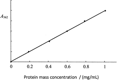
(2) Evenly mix, keep 37 °C for 30min, and then measure the absorbance by colorimetry at the wavelength of 562mm. The standard curve is drawn with the mass concentration of protein (mg/mL) as the abscissa and the absorbance value as the ordinate (Figure 2-1-5).
2. Determination of Protein Content in Samples
Take 100μL of the protein sample to be tested or dilute the corresponding multiple (the concentration should be within the range of the protein standard curve), add 3.0 mL of BCA working solution, mix it well and treat it the same as the standard sample, measure the absorbance value by colorimetry at the wavelength of 562mm, find out the protein concentration according to the standard curve, and then multiply it by the dilution multiple to obtain the true mass concentration of the protein sample to be tested.
1. If the sample contains EDTA, EGTA, DTT, ammonium sulfate and lipid, the test results will be affected, and the high concentration of detergent will also affect the test results. The interfering substances can be removed by TCA precipitation.
2. After adding BCA working solution, it can also be placed at room temperature for 2h or at 60 °C for 30min. When the protein concentration is determined by BCA method, the absorbance value will increase with the extension of time, and the color reaction will be accelerated with the increase of temperature. If the concentration is low, it is suitable to incubate at a higher temperature or extend the incubation time.
3. The mass concentration of the sample to be tested has a good linear relationship within the concentration range of 10-2000μL /mL.
* 1 This can be distilled water, protein buffer solution or salt solution. Depending on the sample dissolution solution.