Protein Interaction (2) Principle and Protocol of EMSA
The electrophoretic mobility shift assay (EMSA), or gel shift assay is a simple and rapid method to detect protein complexes with nucleic acids. EMSA originally used widely in the study of sequence-specific DNA-binding proteins such as transcription factors, has been further developed to investigate DNA-protein interactions, RNA-protein interactions, and even DNA-RNA interactions. It is also applied to qualify and quantify proteins that specifically bind to given nucleotides, enabling to accommodate a wide range of binding conditions.
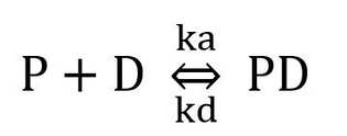
EMSA based on the principle that the rate of migration of the complex of DNA and protein is slower than single DNA fragment or double stranded oligonucleotide during a nondenaturing polyacrylamide gel. Besides, the kinetic analysis of EMSA is another basic theory to underlying the method. The protein (P), binding to a unique site on a DNA (D), will form a complex PD, in equilibrium with the free components:

Where ka is the rate of association and kd is the rate of dissociation. When there is a strong interaction between protein and DNA, Ka>Kd, a distinct band (PD) is observed. However, because of the dissociation occurs during electrophoresis, a faint smear would also show between the two major bands. If a single DNA molecule has multiple binding sites for an individual protein, there will be multiple complexes formed, and we could observe many bands.
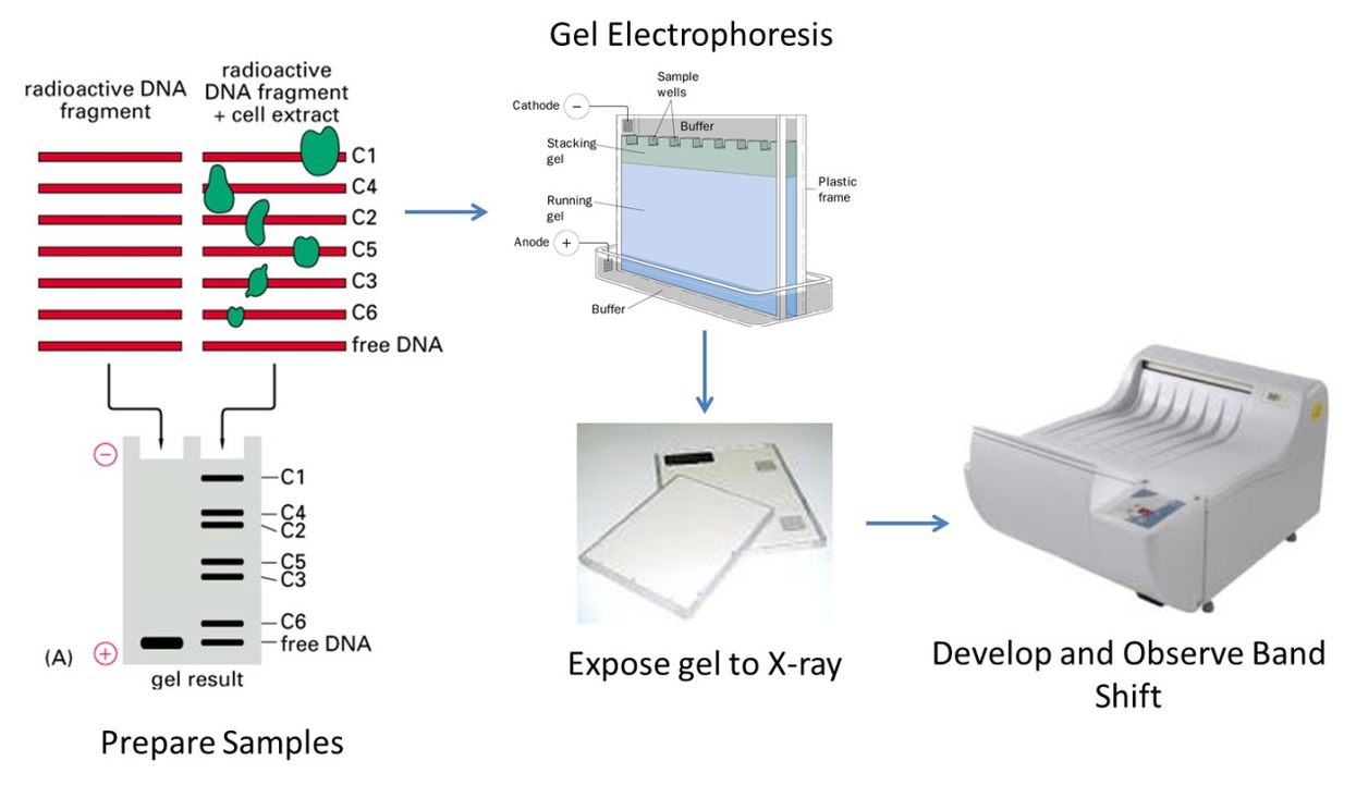
Purified proteins, nuclear or cell extract preparations co-incubate with 32P end-labeled DNA fragment containing the putative protein binding site. Then the reaction products are analyzed on a nondenaturing polyacrylamide gel. The specific binding-protein was determined by competition experiments. Buffers with high ionic strength during electrophoresis can produce electrophoretic bands that are more vivid than the buffer profile with low ionic strength. However, there are some DNA in buffers with high ionic strength cannot interact with protein. Therefore, for each protein-DNA interaction, both buffer systems should be tested.
EMSA is simple to perform, thus it could be used for a wide range of binding conditions. The assay is also highly sensitive within small protein and nucleic acid concentrations. And ranges of nucleic acid sizes and structures are suitable for the assay. Additionally, the EMSA assay words well for both purified proteins and crude cell extracts.
But there are also limitations on EMSA assay. Firstly, weak interaction or rapid dissociation during electrophoresis can prevent detection of complexes brand. Secondly, electrophoretic mobility of a protein-nucleic acid complex depends on many factors other than the size of the protein. Thus we could not measure the protein directly by observing the gel. Thirdly, the result of the assay provides little direct information on the location of the nucleic acid sequences that binding the protein. Finally, the time resolution of the current assay is defined by the interval required for manual solution handling.
 Fig1. EMSA procedure
Procedure:
1. Probe Labeling
- Select restriction enzymes that produce the shortest DNA fragment containing the sequence of inteterest.
- Run a sample on an agarose gel to check the digestion is complete.
- Add CIP and 10×CIP buffer directly to the digestion reaction mix and fill with H2O. Incubate at 37°C for 90 min.
- Incubate the reaction mix at 70°C for 10 min to eliminating and inactivating CIP.
- Precipitate DNA on dry ice for 30min with1/10 volume of NaOAc 3 M pH 5.2 and 2 volumes of cold 90% ethanol.
- Centrifuge for 15 min at 4°C.
- Resuspend DNA in 33 mL of H2O and add 5 mL of 10×kinase buffer, 2 mL of T4 polynucleotide kinase, and 100 mCiof [g-32P] ATP. Mix and incubate at 37°C for 2 h.
- If needed, precipitate DNA and digest the remainder with the second restriction enzyme following the same procedure.
2. Probe Isolation
- Clean and dry the polyacrylamide gel apparatus and its accessories.
- Prepare the 6% polyacrylamide gel.
- Mount the gel in the electrophoresis tank and fill the chamber with 1× TBE.
- Add 6×loading buffer to the digested sample and load into two wells. Migration should be stopped when bromophenol blue reaches two-third of the gel length.
- Under radioactive protection, disassemble the apparatus. Mark the gel and labeled bands in a dark room. Using a razor blade cut the bands interested.
- Place the acrylamide fragment in a dialysis tubing and add 1 mL of 1× TBE, place the dialysis tubing in a standard electrophoresis tank for agarose gel. Run for 15min.
- Using a Pasteur pipette, transfer the labeled probe-containing TBE from the dialysis tubing into microcentrifuge tubes.
- Precipitate DNA on dry ice for 30min with1/10 volume of NaOAc 3 M pH 5.2 and 2 volumes of cold 90% ethanol. Centrifuge for 15 min at 4°C.
- Resuspend labeled DNA in order to obtain 30,000 cpm/mL.
3. EMSA
- Clean and dry the polyacrylamide gel apparatus and its accessories.
- Prepare the 5-15% polyacrylamide gel according the binding protein.
- Pre-electrophoresis at approximately 10 V cm–1 of gel length. Reduce this voltage if gel heating is evident.
- Prepare samples and equilibrate for 30 min at 20 °C ± 1°C
- Add 6×loading buffer to the digested sample and load into two wells. Migration should be stopped when bromophenol blue reaches two-third of the gel length.
- Under radioactive protection, disassemble the apparatus, leaving the gel adherent to one of the plates.
- Gel autoradiography in dark room. Place film or phosphor screen in an exposure cassette. Place the wrapped gel and plate assembly in the cassette, with gel-side toward film or screen. Close the cassette. Expose film or screen at 4 °C for intervals up to 24 h, and at –80 °C for longer intervals.
Sample Analysis
Example 1:
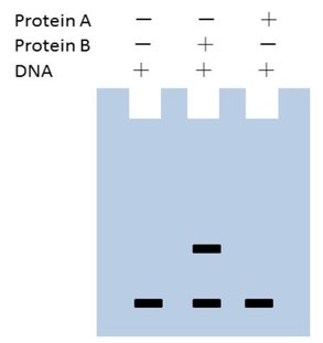
 Fig2. EMSA Result 1
Analysis: Protein B binds to DNA and protein A does not interact with DNA.
Example 2
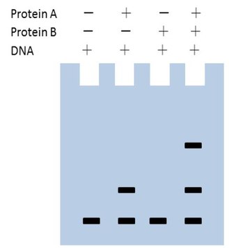
 Fig3. EMSA Result 2
Analysis: Protein A binds to DNA. Protein B binds with DNA if there is protein A. The upper band in column four presents DNA+A+B. Protein B does not interact with DNA independently.

