We use cookies to understand how you use our site and to improve the overall user experience. This includes personalizing content and advertising. Read our Privacy Policy
🧪 MCF-7-006HCL
Source:
Species: Human
Tag:
Conjugation:
Protein Length:
🧪 A431-007HCL
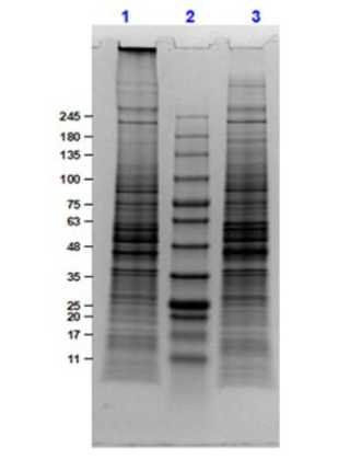
🧪 HEK293-008HCL
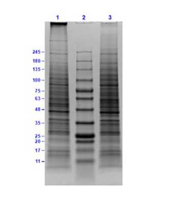
🧪 Raji-009HCL
🧪 Jurkat-010HCL
🧪 A549-011HCL
🧪 U-2OS-012HCL
🧪 Foreskin Fibroblast-013HCL
🧪 HEK293-014HCL
🧪 PC-3-015HCL
🧪 U-87-016HCL
🧪 U-251-017HCL
🧪 Daudi-018HCL
🧪 A549-019HCL
🧪 K-562-020HCL
Inquiry Basket
Compare Basket

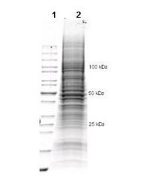
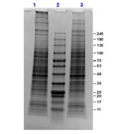
![SDS-Page of HEK239 Whole Cell Lysate.<br />Lane 1: Reduced HEK239 Whole Cell Lysate [10 μg].<br />Lane 2: Opal Prestained Molecular Weight Marker.<br />Lane 3: Non reduced HEK239 Whole Cell Lysate [10 μg].<br />4-20% SDS-PAGE gel, Coomassie Stained.](/productimages/HEK293-008HCL, 1.jpg)
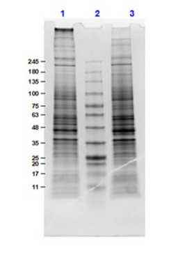
![SDS PAGE Results of Raji Whole Cell Lysate.<br />Lane 1: Raji Whole Cell Lysate Reduced [10 μg].<br />Lane 2: Opal Prestained Molecular Weight Marker.<br />Lane 3: Raji Whole Cell Lysate Non-Reduced [10 μg].<br />4-20% Gel, Coomassie Stained.](/productimages/Raji-009HCL, 1.jpg)

![Western Blot of Human MAPK1 (ERK2) Knockout A549 Cell Lysate.<br />Lane 1: Opal Prestained MW Marker.<br />Lane 2: A549 WCL Parental.<br />Lane 3: A549 MAPK1 KO Clone 4.<br />Lane 4: A549 MAPK1 KO Clone 10.<br />Lane 5: A549 MAPK1 KO Clone 15.<br />Lane 6: HeLa WCL Parental<br />Load: 10 μg lysate/lane. <br />Primary Antibody [Blot A] Anti-MAPK1(ERK2) ~44 kDa; [Blot B] stripped and re-probed with Anti Tubulin ~50 kDa; at 1 μg/mL overnight at 2-8 centigrade.<br />Secondary Antibody: Goat Anti-Rabbit IgG HRP at 1:30,000 for 1hr at RT.<br />Block: BlockOut Buffer for 1 hr at RT.<br />No detection of expected band at ~44 kDa is observed in MAPK1 (ERK2) knockout A549 when compared with unmodified A549 cell lysates by Western blot.](/productimages/A549-019HCL, 1.jpg)
