
Signaling Kits


Background
Overview
Signaling Kits, or signal transduction kits, are tools specifically designed to study intracellular signaling pathways. These kits typically contain reagents and tools for detecting the activity of specific signaling molecules (such as kinases, phosphatases, transcription factors, etc.), as well as biomarkers and detection systems for analyzing signal transduction events.
Product Advantages
Safety and environmental protection: Many signal transduction kits use non-isotopic chemiluminescence detection systems to avoid the use of radioactive substances, reducing environmental pollution and experimental risks.
High sensitivity and specificity: The reagents and antibodies in the kit are optimized to provide highly sensitive and specific signal detection with reduced background interference.
Ease of operation: The design of the kit aims to simplify the experimental process and provide detailed operating instructions, making the experimental operation more convenient and fast.
Classification of Signaling Kits
Signaling Pathway Analysis Kits are used to study the activation status of specific signal transduction pathways, such as MAPK, PI3K/AKT, JAK/STAT, etc.These kits are the hottest and most widely covered category of all kinds.
Intracellular Signaling Assays are used to detect the activity of intracellular signaling molecules, such as kinases, phosphatases, and transcription factors.
Flow Cytometry Signaling Kits are used to study cell surface and intracellular signaling molecules using flow cytometry techniques. These kits typically contain antibodies coupled with different fluorescent dyes to detect the expression and activation status of specific signaling molecules.
Enzyme-Linked Immunosorbent Assay Kits (ELISA) are used to quantitatively detect specific proteins or their post-translational modifications in cell culture supernatants or biological samples.
The Chromatin Immunoprecipitation kits are used to investigate protein-DNA interactions and the influence of epigenetic modification on gene expression.

Meanings of Signaling Pathway Study
Regulation of cell function. Signaling pathways are the pathways through which information is transmitted within cells, and they allow cells to make precise responses to various biological signals. By studying these pathways, scientists can understand how cells regulate basic biological processes such as proliferation, differentiation, migration, and apoptosis.
The revelation of the mechanism of disease. The occurrence and development of many diseases, including cancer, autoimmune diseases, neurodegenerative diseases, etc., are associated with abnormal signaling pathways. Studying these pathways will help to reveal the molecular mechanism of diseases and provide new ideas for disease prevention, diagnosis and treatment.
Drug development and target screening. Key molecules in signaling pathways, such as kinases, receptors, and transcription factors, are often targeted for drug development. By studying the signaling pathway, new drug targets can be discovered and validated, and the drug development process can be accelerated.
The realization of individualized medicine. The study of the signaling pathway helps to understand the differences in gene expression and protein activity between individuals, which is of great value for developing individualized medical strategies and making personalized treatment plans.
Understanding intercellular communication. Cells communicate with their surroundings and other cells through signaling pathways. Studying these pathways helps reveal how cells work together in tissues and organs, and how they participate in complex biological systems.
Applications
Signaling Kits, or signal transduction kits, are tools specifically designed to study intracellular signaling pathways. These kits typically contain reagents and tools for detecting the activity of specific signaling molecules (such as kinases, phosphatases, transcription factors, etc.), as well as biomarkers and detection systems for analyzing signal transduction events. Here are some of the main applications and features of signal transduction kits:

Analysis of signal transduction pathways. Signal transduction kits can be used to study how cells respond to external stimuli through various signaling molecules, such as growth factors, cytokines, hormones, etc. Typically includes antibodies, substrates, or inhibitors to detect the activity of specific signaling molecules, as well as reporter gene systems for signal transduction analysis.
Drug screening and development. During drug development, signal transduction kits can be used to screen and evaluate the effects of potential drugs on specific signaling pathways. These kits provide high-throughput screening platforms that help researchers quickly identify small molecule compounds or biologics that affect signaling.
Study of disease mechanism. Abnormal signal transduction is closely related to the occurrence and development of many diseases, and signal transduction kits can be used to study the molecular mechanisms of these diseases. By analyzing changes in disease-related signaling pathways, researchers can better understand the pathogenesis of the disease and provide a basis for the development of treatment strategies.
Cell function research. Signal transduction is the key to cell function regulation, and signal transduction kits can be used to study cell proliferation, differentiation, migration and other biological functions.
Experimental techniques related to signal transduction. Flow cytometry, gel migration or electrophoretic mobility detection (EMSA), ELISA and other techniques are often used in signal transduction studies. Signal transduction kits are often combined with these experimental techniques to provide the necessary reagents and protocols to support accurate experimental operations and data analysis.
Case Study
Case Study 1: Notch1/CSL Reporter Kit (Notch Signaling Pathway) (Kit-1786)
The family of Notch proteins plays a key role in cell fate determination. Additionally, Notch proteins regulate critical functions of the endothelium, as well as other recruited supporting cells, in concert with other pathways. Despite significant advances in the field and extensive studies focused on elucidating this pathway, many questions remain regarding Notch activation and its upstream/downstream effects, with vascular biology constituting one area of particular interest. Here, the researchers provide a brief description of the components and functions of the Notch pathway in vasculature, followed by a detailed compilation of recommended methods of evaluation in vitro and in vivo. They provide a rationale for key elements when choosing different approaches and controls, strengths and limitations, and essential considerations when providing a meaningful interpretation of results. The aim is to describe a careful approach to assessing Notch function in endothelial cells, based on underlying principles, with the overall goal of obtaining physiologically relevant information that will enhance our understanding of this pathway and its role in vascular biology.
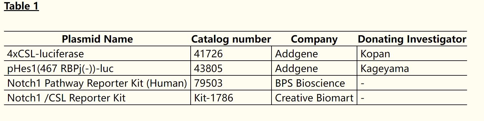
(Lydia L. Wu, 2021)
Fig1. Some luciferase constructs that can be used to detect Notch activation in vitro.
Case Study 2: cAMP Activity Assay Kit
5-demethylnobiletin exhibits a broad spectrum of biological activities such as anti-cancer, anti-inflammatory, cardiovascular protective and neuroprotective effects, however, its effect in melanogenesis remains uninvestigated. The current work aims to confirm the bioactivity and mechanism of 5-demethylnobiletin in stimulating melanogenesis. Multiple biological assays on melanogenesis-associated proteins such as melanin content detection, tyrosinase activity colorimetric assay, cAMP activity assay and Fontana-Masson ammoniacal silver staining were used to confirm the role of 5-demethylnobiletin in stimulating melanin synthesis and the transportation of melanosomes. As confirmed by multiple biological assays, 5-demethylnobiletin is found to stimulate dendrite structure formation in cells, melanin synthesis and the transportation of melanosomes, via inducing the phosphorylation of cAMP response element-binding protein (CREB) and increasing the intracellular levels of cAMP in vitro through the PKA-dependent pathway.
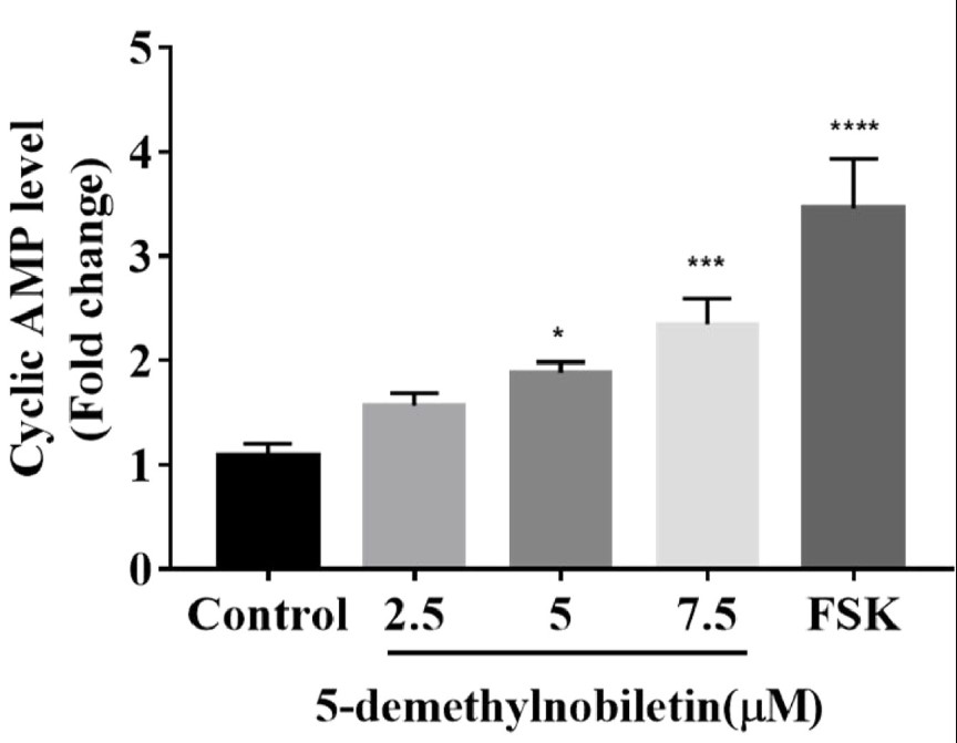
(Hui Miao Wang, 2022)
Fig2. B16F10 cells were incubated with 5-demethylnobiletin (2.5, 5 and 7.5 μM) for 30 min, and the changes of intracellular cAMP level were analyzed by using a cyclic AMP competitive Kit.
Case Study 3: Cell Transformation Assay Kit
This study focused on understanding matrix adhesion pathways that control the oral carcinoma response to cisplatin. The studies revealed that adhesion of HN12 and JHU012 oral carcinomas to carcinoma matrix supported tumor cell proliferation in response to treatment with cisplatin. Disruption of talin expression in HN12 cells adherent to carcinoma matrix increased cisplatin induced proliferation. Pharmacological inhibitors were used to determine signaling events required for talin deficiency to regulate cisplatin induced proliferation. Pharmacological inhibition of NF-kB reduced proliferation of talin-deficient HN12 cells treated with 30 µM cisplatin. Nuclear NF-kB activity was assayed in HN12 cells using a luciferase reporter of NF-kB transcriptional activity. Nuclear NF-kB activity was similar in HN12 cells adherent to carcinoma matrix and collagen I when treated with vehicle DMSO. Following treatment with 30 µM cisplatin, NF-kB activity is maintained in cells adherent to carcinoma matrix whereas NF-kB activity is reduced in collagen I adherent cells. Talin overexpression was sufficient to trigger NF-kB activity following treatment with cisplatin in carcinoma matrix adherent HN12 cells in a process disrupted by FAK siRNA.
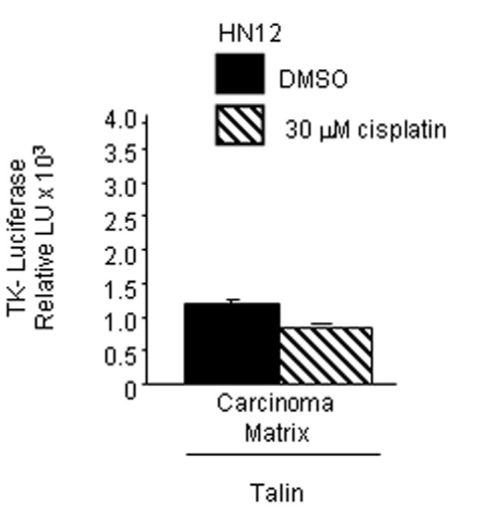
(Karen E Eberle, 2011)
Fig3. HN12 cells transfected with talin cDNA expression vector together with tk-luciferase (lacking the NF-kB regulatory element) and renilla luciferase adherent to carcinoma matrix were treated with DMSO and 30 µM cisplatin.
Advantages
- Product diversity. Our extensive product line includes signaling kits for different signaling pathways such as Notch Signaling Pathway, JNK Pathway, cAMP/PKA Signaling Pathway, PI3K/AKT Pathway, Myc Signaling Pathway and so on.
- Technical specialization. Our R&D team are dedicated to developing all kinds of test kits. They have a rich academic background ranging from Molecular biology, Pharmacy to Cell biology, Medicine.
- Customized service. We not only provide standardized test kits, but also provide customized test kits according to customer needs. The flexibility of this service can better meet specific research needs and provide customers with more personalized solutions.
Creative BioMart offers a series of Signaling Kits for help to study the activation status of specific signal transduction pathways, such as MAPK, PI3K/AKT, JAK/STAT, etc. You can also let us know if you have any customized requirements. Please contact us for more product details.
References
- WU, Lydia L. et al. The long and winding road: detecting and quantifying Notch activation in endothelial cells. Vascular Cell. 2021;13(1), 1.
- Wang HM.; et al. Natural Citrus flavanone 5-demethylnobiletin stimulates melanogenesis through the activation of cAMP/CREB pathway in B16F10 cells. Phytomedicine. 2022;98:153941.
- Eberle KE.; et al. Carcinoma matrix controls resistance to cisplatin through talin regulation of NF-kB. PLoS One. 2011;6(6):e21496.