


Tumor Markers
🧪 HB-42P
Source: Pig
Species: Pig
Tag: Non
Conjugation:
Protein Length:

🧪 HB-43R
Source: Rat
Species: Rat
Tag: Non
Conjugation:
Protein Length:

🧪 HB-44R
Source: Rabbit
Species: Rabbit
Tag: Non
Conjugation:
Protein Length:

🧪 HB-45R
Source: Simian
Species: Simian
Tag: Non
Conjugation:
Protein Length:

🧪 INHBE-797H
Source: E.coli
Species: Human
Tag: His
Conjugation:
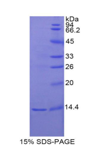
Protein Length: Thr237~Ser350
🧪 Inhbe-798M
Source: E.coli
Species: Mouse
Tag: GST&His
Conjugation:
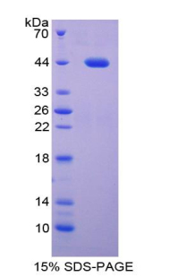
Protein Length: Ser102~Val344
🧪 Inhbe-799R
Source: E.coli
Species: Rat
Tag: His
Conjugation:
Protein Length: Thr237~Ser350


🧪 CALCA-794C
Source: E.coli
Species: Chicken
Tag: His
Conjugation:
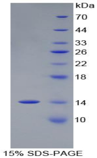
Protein Length: Ala26~Asn138
🧪 Calca-795M
Source: E.coli
Species: Mouse
Tag: GST&His
Conjugation:
Protein Length: Val26~Asn136
What are Tumor Markers?
Tumor markers are substances, often proteins, which are produced by the body in response to cancer growth or by the cancer tissue itself. These markers can be found in the blood, urine, stool, tumors, or other tissues and bodily fluids of some patients with cancer. They are used to help diagnose, predict, and monitor the progression of certain types of cancers.
Tumor Marker Tests
Tumor marker tests measure the levels of these substances in the body to assist in the detection and management of cancer. These tests are rarely used alone for cancer diagnosis but can provide valuable information when used in conjunction with other diagnostic methods.
Blood Tests: The most common method to measure tumor markers. A sample of blood is drawn and analyzed for the presence and concentration of specific markers.
Urine Tests: Some markers can be detected in urine. A sample is collected and analyzed similarly to blood tests.
Tissue Biopsies: Involves removing a piece of tissue from the tumor. The tissue is examined for the presence of tumor markers.
Stool Tests: For certain cancers, markers are measured in stool samples.

How Tumor Marker Tests are done
Sample Collection: Depending on the method, a blood sample may be taken from a vein, urine collected, or a biopsy performed.
Laboratory Analysis: The sample is analyzed in a laboratory setting using various biochemical assays, such as immunoassays or molecular techniques, to detect and quantify the tumor marker.
Understanding the Results
Elevated Levels: High levels of a tumor marker may suggest the presence of cancer, a recurrence of cancer, or a benign condition. However, elevated levels are not definitive for cancer diagnosis.
Normal Levels: Normal levels can indicate that cancer is not present, or that the cancer does not produce that particular marker.
Trend Over Time: More important than a single measurement; regular monitoring can help determine whether a cancer is growing or shrinking in response to treatment.
Tumor marker tests are usually not the sole diagnostic tool but are used in conjunction with imaging studies and medical history to diagnose or monitor cancer. They are more often used to monitor treatment effectiveness or check for recurrence in patients with a known cancer diagnosis.
Types of Tumor Markers and Associated Cancers:
Alpha-fetoprotein (AFP):
Liver cancer
Germ cell tumors
Beta-hCG (human chorionic gonadotropin):
Germ cell tumors
Trophoblastic disease
CA-125 (Cancer Antigen 125): Ovarian cancer
CA 15-3/CA 27.29: Breast cancer
CA 19-9:
Pancreatic cancer
Gallbladder cancer
Bile duct cancer
Gastric cancer
Calcitonin: Medullary thyroid cancer
Carcinoembryonic antigen (CEA): Colorectal cancer, Also elevated in breast, lung, stomach, and pancreatic cancers
Prostate-specific antigen (PSA): Prostate cancer
Lactate dehydrogenase (LDH):
Germ cell tumors
Lymphomas
Neuron-specific enolase (NSE):
Small cell lung cancer
Neuroblastoma
These markers are utilized depending on the suspected type of cancer and are interpreted within the clinical context, including symptoms and imaging studies.
Case Study
Case 1: Winarno GNA, Harsono AB, Suardi D, Salima S, Mantilidewi KI, Bayuaji H, Mulyantari AI, Yulianto FA, Susiarno H. Nomogram development for predicting ovarian tumor malignancy using inflammatory biomarker and CA-125. Sci Rep. 2024 Jul 9;14(1):15790. doi: 10.1038/s41598-024-66509-9. PMID: 38982118; PMCID: PMC11233513.
This study aims to assess the role of pretreatment Neutrophil-to-Lymphocyte Ratio (NLR), Lymphocyte-to-Monocyte-Ratio (LMR), Platelet-to-Lymphocyte Ratio (PLR), and CA-125 in distinguishing benign and malignant ovarian tumors, while also constructing nomogram models for distinguish benign and malignant ovarian tumor using inflammatory biomarkers and CA-125.
 Fig2. Receiver operating characteristic (ROC) curves for inflammatory biomarkers and CA125 in predicting ovarian tumor malignancy. (A) ROC curve for Neutrophil-to-Lymphocyte Ratio (NLR) with an AUC of 0.7051. (B) ROC curve for Lymphocyte-to-Monocyte Ratio (LMR) with an AUC of 0.3386. (C) ROC curve for Platelet-to-Lymphocyte Ratio (PLR) with an AUC of 0.7069. (D) ROC curve for Cancer Antigen 125 (CA-125) with an AUC of 0.7314.
Case 2: Lung Cancer Cohort Consortium (LC3). The blood proteome of imminent lung cancer diagnosis. Nat Commun. 2023 Jun 1;14(1):3042. doi: 10.1038/s41467-023-37979-8. PMID: 37264016; PMCID: PMC10235023.
The authors measured between 392 and 1,162 proteins in blood samples drawn at most three years before diagnosis in 731 smoking-matched case-control sets nested within six prospective cohorts from the US, Europe, Singapore, and Australia. They identify 36 proteins with independently reproducible associations with risk of imminent lung cancer diagnosis (all p < 4 × 10⁻⁵).
 Fig3. Biological context of the 36 proteins associated with risk of imminent lung cancer diagnosis. Relationship between our 36 proteins and the 10 hallmarks of cancer described by Hanahan and Weinberg, based on their descriptions and functions available on GeneCards, the Human Protein Atlas, and Uniprot. Each hallmark is represented by a different color.
Related Resource
- Apoptosis in Cancer
- Autophagy
- Cancer Biomarkers
- Cancer Drug Targets
- Cancer Immunology
- Cell Cycle
- Cell Metabolism
- Oncoproteins
- Signal Transduction in Cancer
- Tumor Microenvironment
- Tumor Suppressors
- PROTAC Targets
- Targets of CAR-T Cell Therapy
- Labeled Car-T Proteins
- ADC Target Protein
- Cell and Gene Therapy