


Co-stimulatory TNF Superfamily Molecules
Related Symbol Search List
- CD40
- TNFSF9
- HVEM
- CD27
- CD40 Ligand
- CD70
- TNFRSF13B
- TNFRSF13C
- TNFRSF4
- TNFRSF8
- TNFRSF9
- TNFSF9
- BAFF
- LIGHT
- TNFSF8
Immunology Background
Background
About Co-stimulatory TNF Superfamily Molecules
The TNF (tumor necrosis factor) superfamily is a group of cytokines and cell surface receptors that play crucial roles in immune regulation, inflammation, cell survival, and apoptosis (programmed cell death). The superfamily is named after one of its founding members, TNF-alpha, which was initially discovered for its ability to induce tumor necrosis (cell death) in certain cancers. The TNF superfamily is characterized by the presence of a conserved structural motif called the TNF homology domain.
The TNF superfamily consists of both soluble cytokines and transmembrane receptors. Soluble cytokines are secreted proteins that bind to specific receptors on the cell surface, while transmembrane receptors are themselves cell surface proteins that can transmit signals into the cell upon ligand binding. These cytokines and receptors function through various signaling pathways to regulate immune responses and maintain tissue homeostasis.
The TNF superfamily members and their receptors have been implicated in numerous physiological and pathological processes, including immune responses, inflammation, autoimmune diseases, cancer, and neurodegenerative disorders. Dysregulation of TNF superfamily signaling can contribute to the development of various diseases.
Targeting the TNF superfamily has therapeutic implications, and drugs that modulate TNF superfamily signaling, such as TNF inhibitors, are used in the treatment of autoimmune diseases like rheumatoid arthritis and inflammatory bowel disease.
Structure of TNF Superfamily Molecules
The TNF superfamily molecules share a common structural motif known as the TNF homology domain (THD) or TNF fold. This structural motif is characterized by a compact, trimeric assembly composed of three identical or similar subunits. Each subunit typically consists of two antiparallel beta-sheets and several alpha-helices.
The overall structure of TNF superfamily molecules can be described as follows:
- Trimeric Assembly: TNF superfamily molecules form homotrimers or heterotrimers, where three identical or similar subunits come together to form a stable trimeric structure.
- Beta-Sheet Core: The TNF fold is primarily composed of two antiparallel beta-sheets arranged in a sandwich-like fashion. The beta-sheets are connected by loops and helices.
- Receptor Binding Site: TNF superfamily molecules possess a receptor binding site located on the surface of the trimer. This site interacts with specific receptors on target cells, triggering downstream signaling events.
- Disulfide Bonds: Disulfide bonds play a crucial role in stabilizing the trimeric structure of TNF superfamily molecules. These covalent bonds form between cysteine residues within each subunit.
- Loop Regions: Loop regions connecting the beta-strands and alpha-helices contribute to the overall stability and flexibility of the TNF superfamily molecules. These loop regions can exhibit structural variations among different family members.
- Post-Translational Modifications: TNF superfamily molecules can undergo various post-translational modifications, such as glycosylation and proteolytic cleavage, which can impact their stability, activity, and receptor binding properties.
It's important to note that while the general structure and trimeric assembly are shared among TNF superfamily members, there can be structural variations and unique features specific to individual molecules. These structural characteristics are essential for their functions as signaling molecules and their interactions with receptors to modulate immune responses and cellular processes.
Co-stimulatory and Co-inhibitory Pathways of the TNF Superfamily Molecules
The TNF superfamily molecules are involved in both co-stimulatory and co-inhibitory pathways, playing crucial roles in immune regulation and modulating immune responses. Here are examples of co-stimulatory and co-inhibitory pathways associated with TNF superfamily molecules:
| Pathways | Molecules | ||
|---|---|---|---|
| Co-stimulatory Pathways | CD40-CD40L Pathway |
|
|
| 4-1BB-4-1BBL Pathway |
|
||
| OX40-OX40 Ligand Pathway |
|
||
| Co-inhibitory Pathways | CTLA-4 (Cytotoxic T-Lymphocyte Antigen 4) Pathway |
|
|
| PD-1 (Programmed Cell Death Protein 1) Pathway |
|
||
| HVEM-BTLA Pathway |
|
||
These are some examples of co-stimulatory and co-inhibitory pathways associated with TNF superfamily molecules. These pathways play important roles in modulating immune responses, maintaining immune balance, and preventing excessive activation or autoimmunity. The balance between co-stimulatory and co-inhibitory signals is crucial for proper immune regulation and immune homeostasis.
TNF Superfamily Molecules
The TNF superfamily is a diverse group of cytokines and receptors that play important roles in immune regulation, inflammation, and cell survival. Here is a list of various TNF superfamily molecules:
| Key molecules | Functions |
|---|---|
| TNF-alpha (TNF-α) |
|
| Fas Ligand (FasL, CD95L) |
|
| TNF-Related Apoptosis-Inducing Ligand (TRAIL) |
|
| CD40 and CD40 Ligand |
|
| TNFSF9 and TNFRSF9 |
|
| HVEM and LIGHT |
|
| CD27 and CD70 |
|
| TNFRSF4 and OX40 Ligand |
|
| TNFRSF8 and TNFSF8 |
|
| BAFF and TNFRSF13C |
|
Role of TNF Superfamily Molecules in Different Diseases
TNF superfamily molecules play critical roles in various diseases due to their involvement in immune regulation, inflammation, cell survival, and tissue homeostasis. Here are some examples of diseases where TNF superfamily molecules have been implicated:
Autoimmune Diseases
- Rheumatoid Arthritis: TNF-alpha is a major driver of inflammation in rheumatoid arthritis. It promotes synovial inflammation, cartilage destruction, and bone erosion.
- Systemic Lupus Erythematosus (SLE): Dysregulated TNF superfamily signaling has been associated with SLE pathogenesis, including abnormal B cell activation and autoantibody production.
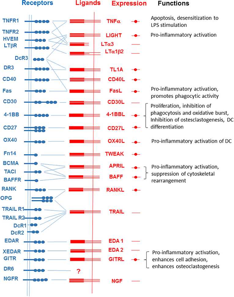 Fig.1 Interactions among members of the TNFSF and TNFRSF. (Lee WH, et al., 2019)
Cancer
- Lymphomas: Aberrant expression of TNF superfamily members, such as CD30 (TNFRSF8) in Hodgkin lymphoma or CD40 in non-Hodgkin lymphoma, contributes to lymphoma development and progression.
- Melanoma: TNF superfamily molecules, including TNF-α and FasL, can influence melanoma cell survival, apoptosis resistance, and immune evasion.
Infectious Diseases
- Viral Infections: TNF superfamily molecules are involved in immune responses against viral infections. For example, TNF-alpha plays a crucial role in antiviral defense against hepatitis B and C viruses.
- HIV/AIDS: Dysregulated expression of TNF superfamily members, such as TNF-alpha, contributes to chronic immune activation, HIV replication, and disease progression.
Inflammatory Bowel Disease (IBD)
- Crohn's Disease and Ulcerative Colitis: TNF-alpha is a key player in the pathogenesis of IBD. Its overproduction leads to chronic inflammation in the gastrointestinal tract, contributing to tissue damage.
Neurological Disorders
- Multiple Sclerosis (MS): TNF-alpha and other TNF superfamily molecules are implicated in the pathogenesis of MS. They contribute to neuroinflammation, demyelination, and neuronal damage.
- Alzheimer's Disease (AD): Dysregulated TNF superfamily signaling, particularly involving TNFRSF1A (TNF receptor superfamily member 1A), has been associated with neuroinflammation and AD pathology.
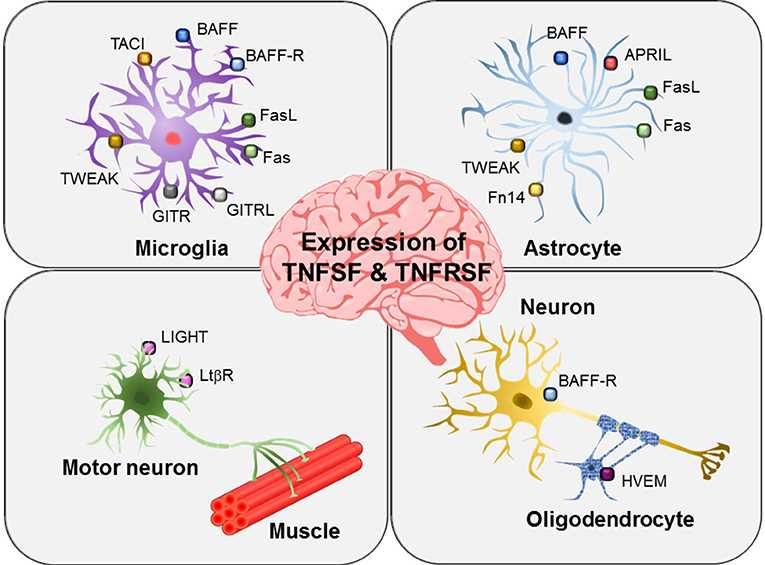 Fig.2 Expression of TNFSF and TNFSRSF members in brain glial cells and neurons. (Lee WH, et al., 2019)
Allergic Diseases
- Asthma: TNF superfamily molecules have been implicated in asthma pathogenesis, contributing to airway inflammation, bronchial hyperresponsiveness, and remodeling.
These examples highlight the diverse roles of TNF superfamily molecules in different diseases. Modulating their activity or targeting specific members of the TNF superfamily has emerged as a therapeutic strategy for several conditions, leading to the development of targeted therapies such as TNF inhibitors for autoimmune diseases. However, the complex and context-dependent nature of TNF superfamily signaling requires further research to fully understand their roles in specific diseases and to develop effective treatments.
Case Study
Case 1: Nguyen J, Pettmann J, Kruger P, Dushek O. Quantitative contributions of TNF receptor superfamily members to CD8+ T-cell responses. Mol Syst Biol. 2021;17(11):e10560.
An operational model inferred by the authors from the data predicts synergy between CD27 and 4-1BB. This prediction is based on the inference that CD27 integrates its signaling into the positive feedback that drives 4-1BB surface expression (A). Thus, the model suggests that CD27 not only directly increases cytokine production, but also increases 4-1BB expression, thereby ameliorating the subsequent co-stimulatory effects induced by 4-1BB engagement. The authors illustrated this by simulating a two-phase stimulation assay (B). These predictions were confirmed experimentally (C and D). Since this synergistic effect is based on activation-induced 4-1BB expression, other activation-induced co-stimulatory receptors should exhibit similar behavior. Indeed, the authors confirmed a similar synergistic effect of GITR, showing that IFN-γ production was significantly enhanced when T cells received early CD27 co-stimulation prior to GITR co-stimulation.
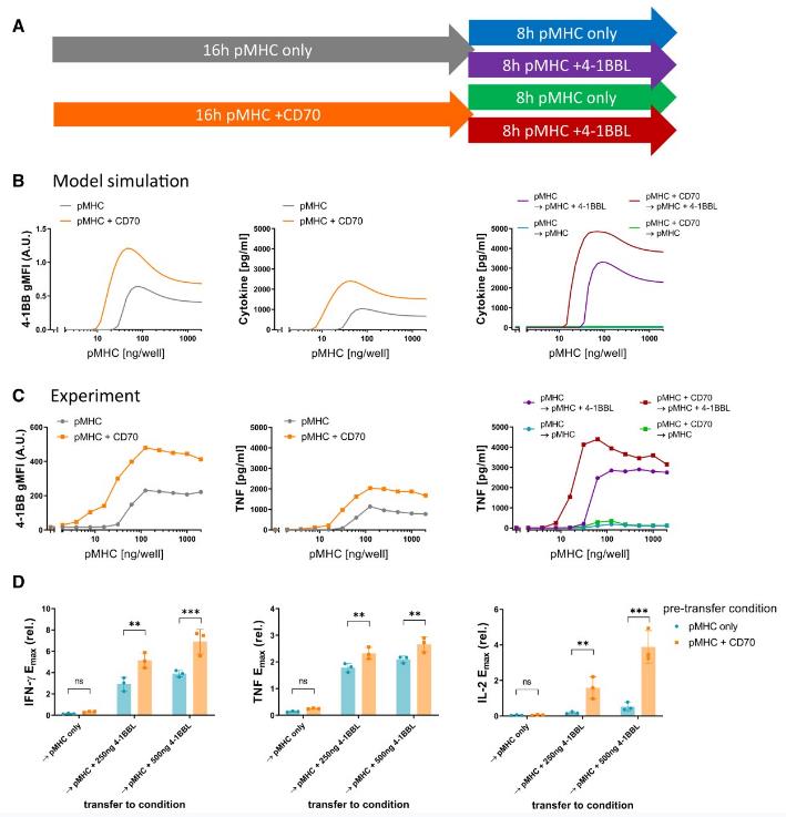 Fig.1 Sequential co-stimulation through CD27 and 4-1BB exhibits synergy.
Case 2: Devarapu SK, Grill JF, Xie J, et al. Tumor necrosis factor superfamily ligand mRNA expression profiles differ between humans and mice during homeostasis and between various murine kidney injuries. J Biomed Sci. 2017;24(1):77.
TNFSF ligands play an important role in the pathogenesis of immune complex organ damage. Therefore, the authors investigated changes in their mRNA expression levels during immune complex glomerulonephritis. The authors used an autologous anti-GBM nephritis animal model and examined TNFSF ligand expression at days 14, 21, and 42. Immune complex glomerulonephritis is associated with progressive increase in glomeruli with integral lesions and infiltration of interstitial F4/80+ macrophages and CD3+ T cells (a). In contrast to IRI and oxalate-induced CKD, mRNA expression of LT-β was maintained at low levels at all time points during progression of immune complex glomerulonephritis (b-c). In conclusion, the authors observed significant differences in renal remodeling induced by persistent crystal injury and immune complex-mediated injury.
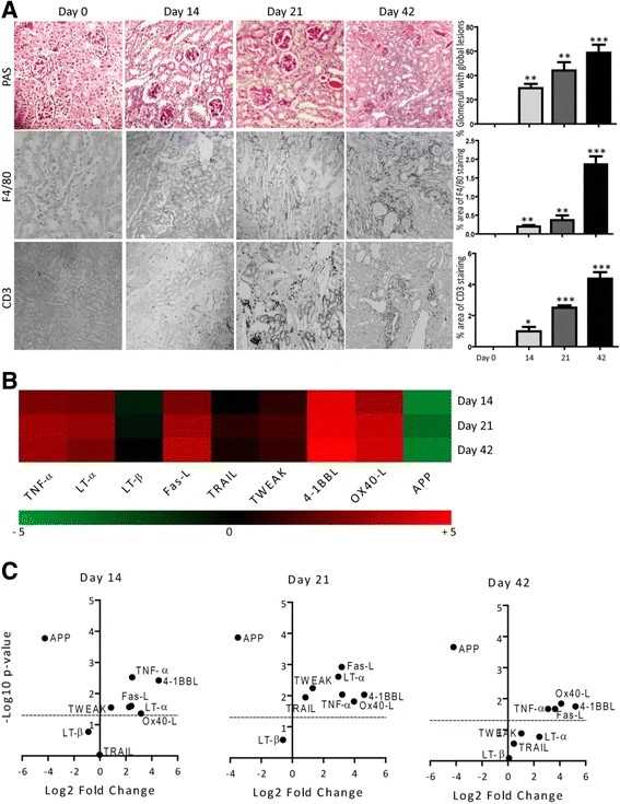 Fig.2 TNFSF ligands mRNA expressions in murine autologous anti-GBM nephritis.
References
- Lee WH, Seo D, Lim SG, Suk K. Reverse Signaling of Tumor Necrosis Factor Superfamily Proteins in Macrophages and Microglia: Superfamily Portrait in the Neuroimmune Interface. Front Immunol. 2019;10:262.
- Bodmer JL, Schneider P, Tschopp J. The molecular architecture of the TNF superfamily. Trends Biochem Sci. 2002;27(1):19-26.
- Cruceriu D, Baldasici O, Balacescu O, Berindan-Neagoe I. The dual role of tumor necrosis factor-alpha (TNF-α) in breast cancer: molecular insights and therapeutic approaches. Cell Oncol (Dordr). 2020;43(1):1-18.