FGA
-
Official Full Name
fibrinogen alpha chain -
Overview
The protein encoded by this gene is the alpha component of fibrinogen, a blood-borne glycoprotein comprised of three pairs of nonidentical polypeptide chains. Following vascular injury, fibrinogen is cleaved by thrombin to form fibrin which is the most abundant component of blood clots. In addition, various cleavage products of fibrinogen and fibrin regulate cell adhesion and spreading, display vasoconstrictor and chemotactic activities, and are mitogens for several cell types. Mutations in this gene lead to several disorders, including dysfibrinogenemia, hypofibrinogenemia, afibrinogenemia and renal amyloidosis. Alternative splicing results in two isoforms which vary in the carboxy-terminus. -
Synonyms
Fibrinogen;FGA;FGA protein;FGB;FGG;Fib2;Fibrinogen A alpha polypeptide;Fibrinogen A alpha polypeptide chain;Fibrinogen alpha chain;Fibrinogen B alpha polypeptide;Fibrinogen beta chain;Fibrinogen G alpha polypeptide;Fibrinogen gamma chain;MGC104327;MGC119422;MGC119423;MGC119425;MGC120405
Recombinant Proteins
- Mouse
- Human
- Cattle
- Rat
- Chicken
- Zebrafish
- Bovine
- Dog
- Goat
- Rhesus macaque
- Rabbit
- Plasma
- Human Plasma
- E.coli
- Mammalian Cells
- Rat plasma
- Protein Conjugation
- HEK293
- Bovine Plasma
- Dog Plasma
- Wheat Germ
- In Vitro Cell Free System
- Yeast
- Baculovirus
- Mammalian cells
- Rabbit Plasma
- Non
- His
- T7
- Avi
- Fc
- GST
Background

Fig1. 3D model representation of the fibrinogen protein. (Rosanna Asselta, 2020)
What is FGA Protein?
FGA gene (fibrinogen alpha chain) is a protein coding gene which situated on the long arm of chromosome 4 at locus 4q31. This gene encodes the alpha subunit of the coagulation factor fibrinogen, which is a component of the blood clot. Following vascular injury, the encoded preproprotein is proteolytically processed by thrombin during the conversion of fibrinogen to fibrin. Mutations in this gene lead to several disorders, including dysfibrinogenemia, hypofibrinogenemia, afibrinogenemia and renal amyloidosis. The FGA protein is consisted of 866 amino acids and FGA molecular weight is approximately 95.0 kDa.
What is the Function of FGA Protein?
FGA protein is one of the key components of fibrinogen in plasma. Fibrinogen is a clotting protein, which is mainly synthesized by liver cells and is the most abundant clotting factor in plasma. The FGA protein plays an important role in blood clotting. When tissues and blood vessels are damaged, fibrinogen is converted by thrombin into water-insoluble fibrin monomers, which are covalent cross-linked to form a stable fibrin web that eventually forms blood clots and stops excessive bleeding. In addition, the alpha chain of fibrinogen contains cell adhesion molecules that promote platelet aggregation and participate in the formation of extracellular matrix.
FGA Related Signaling Pathway
In endometriosis, FGA activates the FAK (plaque adhesion kinase) /AKT (protein kinase B) /MMP-2 (matrix metallopeptidase 2) signaling pathway by binding to integrins via the arginyl-glycinyl-aspartate (RGD) sequence, thereby enhancing the migration and invasion capacity of human endometrial stromal cells. In lung cancer, loss of FGA promotes tumor growth and metastasis by activating the integrin-Akt signaling pathway. In gastric cancer, FGA regulates the FAK/ERK (extracellular signal-regulated kinase) signaling pathway by inhibiting ITGA5 (integrin α5), inhibiting tumor metastasis and inducing autophagic cell death.
FGA Related Diseases
FGA is a key plasma protein component whose abnormality or loss of function is associated with a variety of diseases, including but not limited to blood clotting disorders, the genetic abnormality fibrinogenemia, an increased risk of thrombosis, and certain types of cancer (such as lung and stomach cancer), where FGA may work through signaling pathways that affect cell migration, invasion, and tumor growth. Promote the development and metastasis of tumors. The abnormal function of FGA may also be related to gynecological diseases such as endometriosis.
Bioapplications of FGA
The applications of FGA are mainly in the medical field, especially in the treatment of blood clotting and thrombosis, such as as an adjunct therapeutic ingredient for hemophilia and other clotting disorders, and in some cases as a biomarker to assess a patient's risk of thrombosis. In addition, functional studies of FGA provide potential targets for the development of targeted therapies for specific cancer treatments, helping to design new drugs and therapies to inhibit tumor growth and metastasis.
Case Study
Case Study 1: Hui Li, 2022
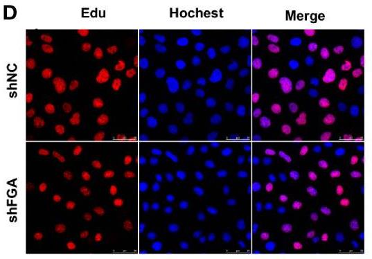
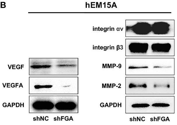
Angiogenesis is the key condition for the development of endometriosis (EM). This study was aimed to elucidate the role of FGA in endometrial stromal cells involved in angiogenesis in EM. The conditioned medium (CM) of human primary eutopic endometrial stromal cells (EuESC) and immortalized endometrial stromal cell line hEM15A with FGA knockdown were collected and used to treat human umbilical vein endothelial cells (HUVECs). Then, tube formation assay, EdU assay, wound assay, transwell assay and flow cytometry assays were performed to assess the function of HUEVCs in vitro. Functionally, CM of endometrial stromal cells with FGA knockdown could inhibit HUEVCs migration and tube formation in vitro and in vivo, while having no significant effect on HUVECs proliferation, apoptosis and cell cycle. Mechanically, the expression of VEGFA, PDGF, FGF-B, VEGF, MMP-2 and MMP-9 was reduced in hEM15A cells with FGA knockdown.

Fig1. Edu assay showing the proliferation of HUEVCs treated with CM from hEM15A shNC and hEM15A shFGA cells.

Fig2. The protein expression of VEGFA, VEGF, MMP-2 and MMP-9 in hEM15A with FGA knockdown was detected by western blotting.
Case Study 2: Guangli Liu, 2022
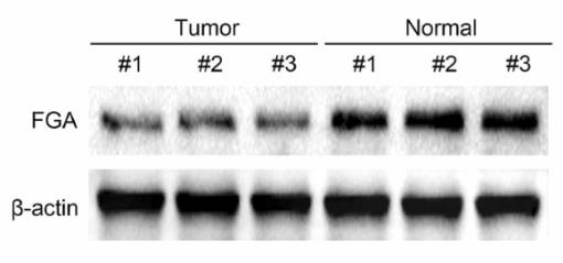
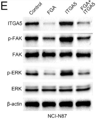
This study aims to investigate the expression levels of fibrinogen α chain (FGA) in human gastric cancer (GC) tissues and cell lines, clarify its role in gastric cancer progression, and explore its underlying mechanism. Luciferase and CHIP assays were performed to confirm the transcriptional regulation of FGA on ITGA5. Immunoblot assays and double-label RFP-GFP-LC3 immunofluorescence analysis were conducted to detect its effects on gastric cancer cell autophagy and FAK/ERK pathway, and in vivo tumor growth assays were further performed. The low expression of FGA in human gastric cancer tissues and cell lines. FGA suppressed gastric cancer cell proliferation, motility, and EMT process, and stimulated cell autophagy. Further, FGA suppressed the expression of Integrin-α5 (ITGA5) and inhibited the FAK/ERK pathway, therefore suppressing the progression of gastric cancer. The in vivo assays further confirmed that FGA suppressed tumor growth of gastric cancer cells in the BALB/c nude mice (18-22 g, female, 8-week-old) through suppressing ITGA5-mediated FAK/ERK pathway in mice.

Fig3. Immunoblot assays showed the expression of FGA in 3 representative gastric cancer tissues and normal tissues.

Fig4. FGA suppresses ITGA5 expression and activates FAK/ERK pathway.
Quality Guarantee
High Purity
.jpg)
Fig1. SDS-PAGE (FGA-2429H)
.
.jpg)
Fig2. SDS-PAGE (FGA-4087H)
Involved Pathway
FGA involved in several pathways and played different roles in them. We selected most pathways FGA participated on our site, such as Complement and coagulation cascades,Platelet activation, which may be useful for your reference. Also, other proteins which involved in the same pathway with FGA were listed below. Creative BioMart supplied nearly all the proteins listed, you can search them on our site.
| Pathway Name | Pathway Related Protein |
|---|---|
| Complement and coagulation cascades | CPB2,C5AR1,MBL1,KLKB1,A2M,C1R,CR2,F9,F10,C6 |
| Platelet activation | PLCB3,PIK3CD,ITPR3,MAPK11,AKT2,PLA2G4F,BTK,RAP1A,MYLK4,PRKG2 |
Protein Function
FGA has several biochemical functions, for example, contributes_to cell adhesion molecule binding,protein binding,protein binding, bridging. Some of the functions are cooperated with other proteins, some of the functions could acted by FGA itself. We selected most functions FGA had, and list some proteins which have the same functions with FGA. You can find most of the proteins on our site.
| Function | Related Protein |
|---|---|
| contributes_to receptor binding | FGB |
| structural molecule activity | COPG,KRT3,FGG,CLDNK,CDC42SE2,EVPL,KRT71,ADD1,SEMG2,SNTG1 |
| protein binding | NOTCH1,SNX20,POC1A,LIMK2,UBE2C,SEPT3,SSTR3,ALDOB,MCM7,PCBD1 |
| protein binding, bridging | CUX1,FLRT3,TCAP,ADAP2,TRIM5,FRMD4A,ST13,SYNM,ANXA1,FLRT2 |
| contributes_to cell adhesion molecule binding | FGB |
Interacting Protein
FGA has direct interactions with proteins and molecules. Those interactions were detected by several methods such as yeast two hybrid, co-IP, pull-down and so on. We selected proteins and molecules interacted with FGA here. Most of them are supplied by our site. Hope this information will be useful for your research of FGA.
FGB;FGG;APOA1;KLK6;PASK;F2;MIS12;KRT18;p29991-pro_0000037941;p29991-pro_0000037946;PRRC2A;d-mannose;crlkekhc;IGHG1;IGHA1;ALB
Resources
Related Services
Related Products
References
- Jobse, A; Pieters, M; et al. The contribution of genetic and environmental factors to changes in total and gamma ' fibrinogen over 5 years. THROMBOSIS RESEARCH 135:703-709(2015).
- Nimje, SV; Panigrahi, SK; et al. Interfacial failure analysis of functionally graded adhesively bonded double supported tee joint of laminated FRP composite plates. INTERNATIONAL JOURNAL OF ADHESION AND ADHESIVES 58:70-79(2015).


