


Active Recombinant Glutathione S-Transferase 
| Cat.No. : | GST-113 |
| Product Overview : | Recombinant Glutathione S-Transferase full length protein (1-224a.a.) expressed in E.coli, having a molecular mass of 26kDa. GST was isolated from an E. colistrain that carries the coding sequence for Schistosoma japonicum GST under the control of a T7 promoter. The GST is purified by proprietary chromatographic techniques. |
- Specification
- Gene Information
- Related Products
- Citation
- Download
| Source : | E.coli |
| Tag : | Non |
| Protein Length : | 1-224 a.a. |
| Description : | Antioxidant enzyme Glutathione S- Transferase (GST) is thought to do the primary cellular defense mechanism against reactive oxygen species. GST reduces lipid hydroperoxides through its Se-independent glutathione peroxidase activity. The enzyme also detoxifies lipid peroxidation end products such as 4-hydroxynonenal (4-HNE). The soluble GST is a 26 kDa protein which occurs as a dimer in all aerobic organisms. Each monomer has two domains, one that binds GSH and is an /-structure similar to thioredoxin and the other, all helical, that binds the hydrophobic substrate. The GST -fusion protein expression system is a widely used recombinant protein expression system that allows a peptide or a regulatory protein domain to be expressed as a fusion to the C-terminus of Schistosoma japonicum GST. Fusion proteins also possess GST -enzymatic activity and can undergo dimerization similar to in vivo. The fusion protein can be purified via GST -affinity column chromatography. In most cases, the desired peptides or domains are removed from GST by applying a specific protease that recognizes and cleaves the linker between the protein domain and GST. The technique has been widely used to generate different kinds of proteins for crystallization, molecular immunology studies, the production of vaccines and studies involving protein-protein and protein-DNA interactions. |
| Form : | Sterile Filtered clear solution. GST supplied in Phosphate Buffered Saline pH 7.4. |
| Bio-activity : | 0.5-2.5 units/mg (please enquire for specific batch value). A unit is defined as the amount of enzyme that conjugate 1.0 u mole of 1-chloro-2,4-dinitrobenzene (CDNB) with reduced glutathione per minute at pH 6.5 at 25C. |
| Molecular Mass : | 26 kDa |
| AA Sequence : | MSPILGYWKI KGLVQPTRLL LEYLEEKYEE HLYERDEGDK WRNKKFELGL EFPNLPYYID GDVKLTQSMA IIRYIADKHN MLGGCPKERA EISMLEGAVL DIRYGVSRIA YSKDFETLKV DFLSKLPEML KMFEDRLCHK TYLNGDHVTH PDFMLYDALD VVLYMDPMCL DAFPKLVCFK KRIEAIPQID KYLKSSKYIA WPLQGWQATF GGGDHPPKSD LVPR. |
| Purity : | Greater than 95% as determined by SDS-PAGE. |
| Applications : | GST can be used for protein-protein interactions assay and protein-DNA interactions assay. |
| Stability : | Store at 4°C if entire vial will be used within 2-4 weeks. Store, frozen at -20°C for longer periods of time. For long term storage it is recommended to add a carrier protein (0.1% HSA or BSA). Avoid multiple freeze-thaw cycles. |
| Full Length : | Full L. |
| ◆ Recombinant Proteins | ||
| GST-2758P | Recombinant Pan-species (General) GST Protein, His-tagged | +Inquiry |
| GST-5835P | Recombinant Plasmodium falciparum GST protein, His-tagged | +Inquiry |
| GST-393 | Recombinant GST Protein | +Inquiry |
| GST-307S | Recombinant Schistosoma japonicum GST Protein | +Inquiry |
| GST-001S | Recombinant Schistosoma japonicum Glutathione S-transferase, BFP-tagged | +Inquiry |
| ◆ Cell & Tissue Lysates | ||
| CPB-382R | Rabbit anti-GST Polyclonal Antibody | +Inquiry |
| CPB-278R | Rabbit Anti-GST Polyclonal Antibody | +Inquiry |
| GST-573SCL | Recombinant Schistosoma japonicum GST cell lysate | +Inquiry |
Insight on a new indolinone derivative as an orally bioavailable lead compound against renal cell carcinoma.
Journal: Bioorganic chemistry PubMed ID: 34020239 Data: 2021/10/22
Authors: Marwa A Fouad, Mayssoune Y Zaki, Walaa R Mahmoud
Article Snippet:The enzyme inhibition assay of compound 4f against VEGFR2, PDGFRα and PDGFR? was performed at the Egyptian company for the production of vaccines, sera, and drugs VACSERA, Giza, EGYPT.The enzyme inhibition assay of compound 4f against VEGFR2, PDGFRα and PDGFR? was performed at the Egyptian company for the production of vaccines, sera, and drugs VACSERA, Giza, EGYPT.. This was carried out using BPS Bioscience? VEGFR2 (KDR) Kinase and PDGFRα (D842I) Assay Kits with PDGFRα (D842I), GST-Tag (BPS Bioscience) or Recombinant Human PDGFR?, GST-tagged (Creative BioMart) according to the manufacturer’s manual.. The assay was carried at concentrations 10–10000 nM for VEGFR2 and PDGFRα, and at 1–1000 nM for PDGFR? using 20 μL of the diluted enzyme (1 ng/μL). (For more information, see Appendix A)The assay was carried at concentrations 10–10000 nM for VEGFR2 and PDGFRα, and at 1–1000 nM for PDGFR? using 20 μL of the diluted enzyme (1 ng/μL). (For more information, see Appendix A)
mTOR direct crosstalk with STAT5 promotes de novo lipid synthesis and induces hepatocellular carcinoma
Journal: Cell Death & Disease PubMed ID: 31409773 Data: 2019/8/14
Authors: Ting Li, Jun Weng, Yi Gao
Article Snippet:Then, the cell lysates were isolated by centrifugation at 12,000 rpm for 10 min in a microcentrifuge, and incubated with anti-mTOR antibody for 2 h at 4 °C, followed by addition of 30 μl of 50% slurry of protein G Sepharose beads for an additional 1 h. Immunoprecipitates were then washed four times with lysis buffer and once with kinase buffer [25 mM HEPES (pH 7.4), 50 mM KCl, 10 mM MgCl2, 250 μM ATP].and incubated with anti-mTOR antibody for 2 h at 4 °C, followed by addition of 30 μl of 50% slurry of protein G Sepharose beads for an additional 1 h. Immunoprecipitates were then washed four times with lysis buffer and once with kinase buffer [25 mM HEPES (pH 7.4), 50 mM KCl, 10 mM MgCl2, 250 μM ATP]. ... In all 0.4 μg of recombinant GST-tagged full-length STAT5 peptide (Creative BioMart, NY, USA) was added to 30 μl kinase buffer.. Reactions were stopped by the addition of 30 μl SDS sample buffer and boiling for 10 min and analyzed by immunoblotting.Reactions were stopped by the addition of 30 μl SDS sample buffer and boiling for 10 min and analyzed by immunoblotting.
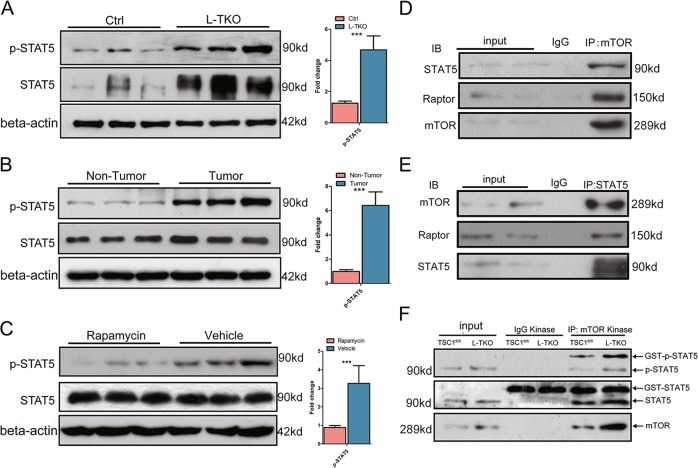
Hepatic p-STAT5 was determined by western blots in L-TKO mice and control littermates ( a ), and in tumor and nontumor areas of L-TKO mice ( b ). c Liver tissues were lysed from rapamycin or vehicle treated 10-month-old L-TKO mice, and p-STAT5 was determined by western blots ( n = 6). Western blots were quantified. *** P < 0.001. d HepG2 cells were immunoprecipitated with anti-mTOR antibody and the amounts of Raptor, and STAT5 co-purified with mTOR were determined by western blot. e HepG2 cells were immunoprecipitated with anti-STAT5 antibody and the amounts of mTOR and Raptor co-purified with STAT5 were determined by western blot. f Cultured hepatocytes cells from Ctrl and L-TKO mice were immunoprecipitated with anti-mTOR antibody and the precipitated mTOR was assayed for kinase activity
Cystic fibrosis transmembrane conductance regulator attaches tumor suppressor PTEN to the membrane and promotes anti Pseudomonas aeruginosa immunity
Journal: Immunity PubMed ID: 29246444 Data: 2018/12/19
Authors: Sebastián A. Riquelme, Benjamin D. Hopkins, Alice Prince
Article Snippet:Chemicals, Peptides, and Recombinant Proteins , , .Chemicals, Peptides, and Recombinant Proteins , , .. Recombinant Human CFTR (1381-1480), GST-tagged , Creative-BioMart , Cat# CFTR-275H Lot # 101018.. Recombinant Human CFTR (1381-1480), GST-tagged , Abnova , Cat# H00001080-Q01 Lot # G7151.Recombinant Human CFTR (1381-1480), GST-tagged , Abnova , Cat# H00001080-Q01 Lot # G7151.
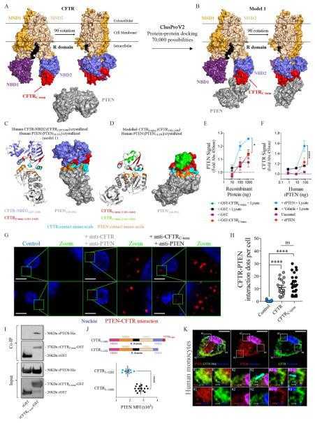
(A) Frontal and Lateral (left) view of the crystallized non-phosphorylated human CFTR and PTEN. PTEN PDB that represents crystalized PTEN does not show the PTEN C-terminus (PTEN14-351), which has been demonstrated as a negative regulator for its interaction with the membrane. Different CFTR domains are shown in different colors (MSD1-2: membrane-spanning domain; NBD1-2: nucleotide-binding domain 1–2; R: regulatory domain). CFTRC-term tail (CFTR1381-1440) is shown in red at the end of NBD2 (red arrow). PTEN (gray) and CFTR-NBD2 PDB structures where submitted to ClusProV2 clustering and modelling. (B) Model 1 (out of 4, see Figure S5A) from ClusProV2 protein-protein interaction modelling showing the interaction between PTEN14-351 and CFTR-NBD2 (CFTR1207-1440). The whole human CFTR structure complexed with human PTEN is showed to better display the orientation of PTEN from the cytoplasm. (C) Human crystallized CFTR-NBD2 and PTEN interaction model 1 from ClusProV2. CFTR1207-1380 is shown in purple. CFTR1381-1440 is shown in red. PTEN is shown in gray. Amino acids from CFTR1207-1440 taking contact with PTEN are displayed in cyan. PTEN amino acids contacting CFTR1207-1440 are shown in orange. (D) I-TASSER modelled CFTR1381-1480 and PTEN interaction model from ClusProV2. CFTR1381-1440 is shown in red. CFTR1441-1480 is shown in green. PTEN is shown in gray. Amino acids from CFTR1381-1480 taking contact with PTEN are displayed in cyan. PTEN amino acids contacting CFTR1381-1480 are shown in orange. (E) ELISA plates were coated either with rGST alone or rGST-CFTR1381-1480. Wells were blocked, washed and incubated with PTEN-containing human 16HBE cell lysates. Plates were revealed for PTEN. (F) ELISA plates were coated either with Vehicle or rPTEN lacking the C-terminus (PTEN1-351). Wells were blocked, washed and incubated with CFTR-containing human 16HBE cell lysates. Plates were revealed for CFTR. (G) CFTR-PTEN and CFTRCterm-PTEN proximity ligation assays (PLA) in human epithelial cells (16HBE). Intracellular detection of CFTR was performed by using either an antibody against a naturally extracellular-exposed loop of CFTR (anti-CFTR) or the CFTRC-term (anti-CFTRC-term). Control cells did not receive primary antibodies. Zoom insets show protein-protein interaction clusters. (H) Quantification of the amount of CFTR-PTEN or CFTRC-term-PTEN clusters obtained with PLA. (I) Pull down analyses between
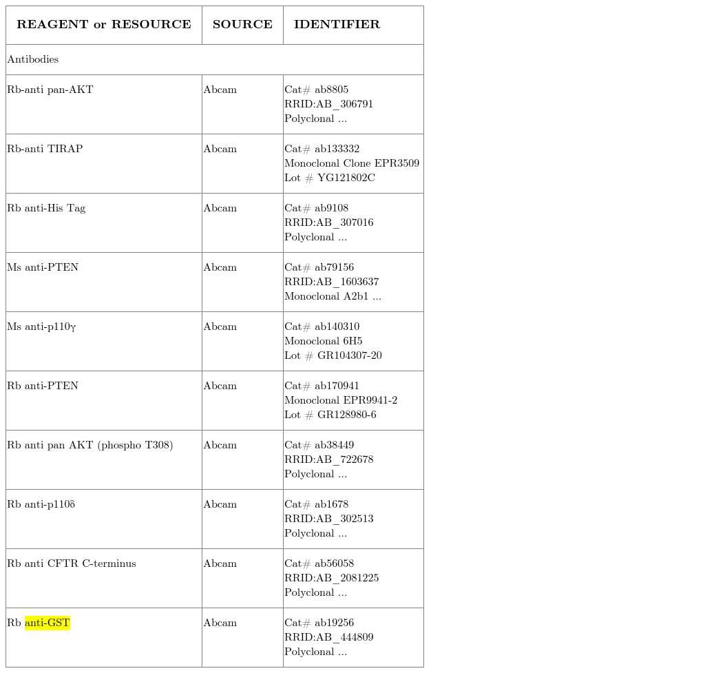
KEY RESOURCES TABLE
TIE2 Associates with Caveolae and Regulates Caveolin-1 To Promote Their Nuclear Translocation
Journal: Molecular and Cellular Biology Data: 2017/11/1
Authors: Mohammad B. Hossain, Rehnuma Shifat, Candelaria Gomez-Manzano
Article Snippet:413 414 In vitro kinase assay.413 414 In vitro kinase assay.. In vitro kinase assays were conducted with 200 ng of purified glutathione 415 S-transferase (GST)–tagged TIE2 active protein (Creative BioMart, no. TEK-7296H) in kinase 416 buffer [25 mM MOPS (pH 7.2), 12.5 mM b-glycerol phosphate, 20 mM MgCl2, 12.5 mM MnCl2, 417 2 mM EDTA (pH 8.0), 5 mM EGTA (pH 7.0), and 0.25 mM DTT] with the addition of 20 mM 418 adenosine triphosphate (ATP).. The substrate, recombinant caveolin-1 protein (NOVUS 419 Biologicals, no. H00000857-P01) was added to the reaction mixture and incubated at 37°C for 420 30 min.The substrate, recombinant caveolin-1 protein (NOVUS 419 Biologicals, no. H00000857-P01) was added to the reaction mixture and incubated at 37°C for 420 30 min.
Not For Human Consumption!
Inquiry
- Reviews (0)
- Q&As (0)


Ask a Question for All GST Products
Required fields are marked with *
My Review for All GST Products
Required fields are marked with *