Mutual regulation between OGT and XIAP to control colon cancer cell growth and invasion
Journal: Cell Death & Disease PubMed ID: 32994395 Data: 2020/9/29
Authors: Hyeon Gyu Seo, Han Byeol Kim, Jin Won Cho
Article Snippet:Recombinant His-Ubiquitin protein and His-UCHL5 were purchased from R&D systems (Minneapolis, MN, USA) and LSBio (Seattle, WA, USA), respectively.Recombinant His-Ubiquitin protein and His-UCHL5 were purchased from R&D systems (Minneapolis, MN, USA) and LSBio (Seattle, WA, USA), respectively.. Recombinant GST-XIAP was purchased from Creative BioMart (Shirley, NY, USA).. Glutathione agarose and Ni-NTA agarose were purchased from Qiagen (Hilden, Germany).Glutathione agarose and Ni-NTA agarose were purchased from Qiagen (Hilden, Germany).
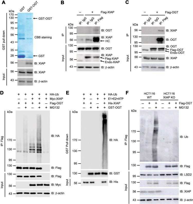
a HCT16 cell lysates were incubated with immobilized recombinant GST-OGT fusion protein (residues 1–1036). GST-OGT was precipitated and the associated XIAP was detected by western blotting with α-XIAP antibodies. GST and GST-OGT were detected by Coomassie brilliant blue staining to verify the amount of protein used in the assay. b HCT116 cells transiently overexpressing Flag-tagged XIAP were immunoprecipitated with α-Flag or IgG control antibodies. Co-immunoprecipitated endogenous OGT was detected by α-OGT antibodies. The same membrane was re-probed with α-XIAP antibodies. Total lysates were blotted with α-OGT and α-XIAP antibodies. c HCT116 cells were transfected with Flag-tagged OGT and immunoprecipitated with α-Flag or IgG control antibodies. Bound endogenous XIAP was detected by α-XIAP antibodies. The same membrane was re-probed with α-OGT antibodies. Total lysates were blotted with α-OGT and α-XIAP antibodies. d OGT in vivo ubiquitination assay. Expression vectors encoding Flag-OGT and HA-ubiquitin (Ub) were transfected into HCT116 cells transiently overexpressing Myc-XIAP as indicated. The cells were treated with 20 μM of MG132 for 4 hours (h) before they were harvested. After being immunoprecipitated with α-Flag antibodies, the ubiquitination of OGT was analyzed by immunoblotting with α-HA antibodies. The same membrane was re-probed with α-Flag antibodies. Equal amounts of total lysates were subjected to immunoblotting with the indicated antibodies. e OGT in vitro ubiquitination assay. GST-OGT was incubated with His-XIAP, HA-Ub, E1, E2, and ATP as indicated. After GST pull-down under denaturing conditions with a buffer containing 2% SDS, the ubiquitination of OGT was analyzed by immunoblotting with α-HA antibodies. The same membrane was re-probed with α-GST antibodies. Equal amounts of XIAP in the reaction were immunoblotted with α-His antibodies. f HCT116 WT or HCT116 XIAP KO cells were transfected with Flag-OGT and treated with 20 μM of MG132 for 4 h. The ubiquitination of immunoprecipitated OGT was analyzed by immunoblotting with α-Ub antibodies. The same membrane was re-probed with α-Flag antibodies. Equal amounts of total lysates were subjected to immunoblotting as indicated. β-actin was used as a loading control. All data are representative of at least three independent experiments.
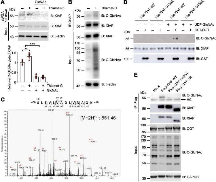
a HCT116 cells were either treated with 1 μM of Thiamet-G for 4 h or not treated with any Thiamet-G. Cell lysates were then subjected to sWGA lectin affinity purification and analyzed with Western blotting for the presence of endogenous XIAP. As a control, 20 mM of the monosaccharide inhibitor GlcNAc was added during sWGA lectin affinity purification. Relative O-GlcNAcylated XIAP levels were plotted ( n = 5 per condition). b HCT116 cells were either treated with 1 μM of Thiamet-G for 4 h or not treated with any Thiamet-G. Immunoprecipitated endogenous XIAP was detected by α-O-GlcNAc antibodies. c Expression vectors encoding Flag-OGT were transiently transfected into HEK293 cells and immunoprecipitated Flag-XIAP was subjected to MS analysis. The CTD MS/MS spectrum of residues 406–419, O-GlcNAcylated XIAP peptides, with the doubly charged precursor ion m/z 851.461426 (M + 2H) 2+ is shown. The b- and y-type product ions were assigned. d Identification of the O-GlcNAc modification sites of XIAP by in vitro glycosylation assay. Purified WT or mutant His-tagged XIAP were used as substrates. O-GlcNAc-modified XIAP was analyzed by α-O-GlcNAc antibodies. The same membrane was re-probed with α-XIAP antibodies. Immunoblotting with α-GST antibodies was conducted to ensure that there was an equal amount of OGT. e Flag-XIAP WT, XIAP S406A, or XIAP ΔRING mutants were transiently overexpressed in HCT116 XIAP KO cells. XIAP WT and XIAP mutants were immunoprecipitated with α-Flag antibodies and blotted with α-O-GlcNAc and α-XIAP antibodies. Equal amounts of total lysates were subjected to immunoblotting with antibodies as indicated. β-actin or GAPDH was used as a loading control. Data are presented as means ± SD of at least three independent experiments. Statistical significance was determined using one-way analysis of variance. * P < 0.05, *** P < 0.001.
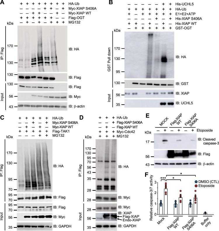
a Expression vectors encoding Flag-OGT and HA-Ub were transfected into HCT116 cells transiently overexpressing Myc-XIAP WT and Myc-XIAP S406A as indicated. Cells were treated with 10 μM of MG132 for 4 h before being harvested. After immunoprecipitation with α-Flag antibodies, the ubiquitination of OGT was analyzed by immunoblotting with α-HA antibodies. The same membrane was re-probed with α-Flag antibody. Equal amounts of total lysates were subjected to immunoblotting with the indicated antibodies. b GST-OGT was incubated with HA-Ub, E1, E2, ATP, His-UCHL5, and either His-XIAP WT or His-XIAP S406A in the presence of UDP-GlcNAc as indicated. After GST pull-down was conducted under denaturing conditions with a buffer containing 2% SDS, the ubiquitination of OGT was detected by immunoblotting with α-HA antibodies. The same membrane was re-probed with α-GST antibodies. The amounts of XIAP and UCHL5 in the reaction were detected with α-XIAP antibody and α-His antibodies, respectively. c Expression vectors encoding Flag-TAK1 and HA-Ub were transiently transfected into HCT116 cells overexpressing Myc-XIAP as indicated. Cells were treated with 10 μM of MG132 for 4 h before being harvested. After immunoprecipitation with α-Flag antibodies, the ubiquitination of TAK1 was analyzed by immunoblotting with α-HA antibodies. The same membrane was re-probed with α-Flag antibodies. Equal amounts of total lysates were subjected to immunoblotting with the indicated antibodies. d Expression vectors encoding Myc-Cdc42 and HA-Ub were transiently transfected into HCT116 cells overexpressing Flag-XIAP as indicated. Before harvest, cells were treated with 20 μM of MG132 for 4 h. After immunoprecipitation with α-Myc antibodies, the ubiquitination of OGT was analyzed by immunoblotting with α-HA antibodies. The same membrane was re-probed with α-Myc antibodies. Equal amounts of total lysates were subjected to immunoblotting with the indicated antibodies. e Expression vectors encoding Flag-XIAP WT and XIAP S406A were transfected into HCT116 cells. Cells were treated with 20 μM of etoposide for 24 h and lysed with NET buffer before being harvested. Activated caspase 3 was monitored with α-cleaved caspase 3 antibodies. Equal amounts of total lysates were subjected to immunoblotting with the indicated antibodies. f HCT116 cells were transfected with Myc-XIAP WT or Myc-XIAP S406A. Cells were treated with 20 μM of etoposide for 24 h and lysed with NET buffer before being harvested. Aliquots containing 50 μg of lysate were added to 100 μM of Ac-DEVD-AFC for measuring caspase-3/7 activity ( n = 6 per condition). Initial rates were analyzed at Ex/Em = 380 / 500 nm. β-actin or GAPDH was used as a loading control. Data are presented as means ± SD of at least three independent experiments. Statistical significance was determined using one-way analysis of variance. * P < 0.05, *** P < 0.001.


