


Myogenic Precursor Markers
Related Symbol Search List
Immunology Background
Overview of Myogenic Precursor Markers
Myogenic precursor cells are early-stage cells, mainly found in the muscles of adult animals and humans, with the potential to further differentiate into mature muscle cells and with the ability to rebuild and repair damaged muscle tissue. Myogenic precursor cell markers are specific protein markers expressed during the differentiation of mesenchymal stem cells (MSCs) to myogenic cells. Researchers have identified a series of specific markers for identifying and isolating these myogenic precursor cells and conducting related studies. Currently, commonly used markers for myogenic precursor cells include M-Cadherin, Pax7, Myf5, MyoD, Myogenin, etc. Of these, Pax7 is a transcription factor that is widely used to mark the embryonic stage of myofibroblasts, while Myf5 and MyoD are expressed in subsequent stages of myogenic precursor cells. Using these markers, researchers can track and characterize myofibroblast precursor cells to gain insight into their developmental processes, mechanisms, and applications in regenerative medicine and tissue engineering.
Research Areas of Myogenic Precursor Markers
- Muscle development and differentiation
Researchers have shed light on the mechanisms of muscle development and differentiation by studying the expression and function of myogenic precursor markers. For example, it was found that Pax7 is widely used to identify and track the presence and differentiation capacity of muscle stem cells. In one study, by analyzing changes in Pax7 expression during mouse muscle development, it was found to be localized in the nucleus of muscle stem cells during the early stages of muscle development and to gradually decrease during muscle development (Relaix et al., 2005).
- Muscle regeneration and repair
By studying these markers, researchers can identify and isolate myogenic precursor cells with regenerative potential, which in turn can facilitate the repair process after muscle injury. For example, it was found that CD56 (also known as NCAM), a myogenic precursor marker, can be used to identify muscle stem cells and precursor cells in the muscle regeneration process. One study used human bone marrow MSCs and used CD56 expression to identify and isolate cell populations with myogenic regenerative potential (Bosnakovski et al., 2008). Researchers are exploring the use of myogenic precursor cell markers to accelerate the muscle regeneration process to promote the natural repair of damaged muscle. By introducing markers, researchers can track the migration, proliferation, and differentiation processes of these myogenic precursor cells after injury. This could help develop more effective strategies for treating skeletal muscle injuries, muscular dystrophy, and muscle diseases in the clinic.
- Muscle disease research
In myotonic dystrophy research, myogenic precursor cells have been found to have a restricted ability to proliferate and differentiate, leading to muscle atrophy and loss of function. A study using a mouse model suggests that myogenic precursor cells in muscular dystrophy patients are inhibited in proliferation and differentiation and may be a key factor in muscle atrophy (Barberi et al., 2007). Muscle atrophy caused by myotonic dystrophy also involves several mechanisms such as inflammatory responses, metabolic disorders, and imbalances in protein metabolism. Researchers have also found that the expression and function of some myogenic precursor markers are altered in myotonic dystrophy. For example, it was found that myotonic dystrophy patients have reduced expression levels of markers such as Pax7 and MyoD in myogenic precursor cells, which may be associated with a decreased ability to regenerate and repair muscle (Penna et al., 2013).
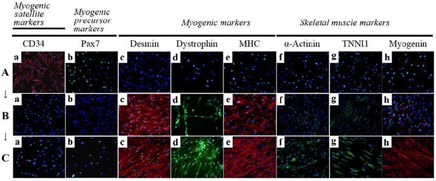 Fig.1 Immunocytochemistry for the detection of myogenic markers. (Park S, et al., 2016)
The study of myogenic precursor markers contributes to our understanding of the process of muscle regeneration and muscle fiber remodeling and also provides a basis for the development of new therapies against muscle diseases and injuries. Further studies will help to reveal the mechanisms of myogenic precursor cell markers in muscle regeneration and functional repair and provide more effective and personalized therapies for clinical applications.
Reference:
- Relaix, F., Rocancourt, D., Mansouri, A., & Buckingham, M. (2005). A Pax3/Pax7-dependent population of skeletal muscle progenitor cells. nature, 435(7044), 948-953.
- Bosnakovski, D., Mizuno, M., Kim, G., Ishiguro, T., Okumura, M., Iwanaga, T., ... & Fukuta-Ohi, H. (2008). Chondrogenic differentiation of bovine bone marrow mesenchymal stem cells (MSCs) in different hydrogels: influence of collagen type II extracellular matrix on MSC chondrogenesis. biotechnology and bioengineering, 99(2), 455-464.
- Barberi, T., Bradbury, M., Dincer, Z., Panagiotakos, G., Socci, N. D., Studer, L., ... & Wagers, A. J. (2007). Derivation of engraftable skeletal myoblasts from human embryonic stem cells. Nature Medicine, 13(5), 642-648.
- Penna, F., Costamagna, D., Pin, F., Camperi, A., Fanzani, A., Chiarpotto, E. M., ... & Costelli, P. (2013). Autophagic degradation contributes to muscle wasting in cancer cachexia. The American journal of pathology, 182(4), 1367-1378.
- Park S, Choi Y, Jung N, et al. Myogenic differentiation potential of human tonsil-derived mesenchymal stem cells and their potential for use to promote skeletal muscle regeneration. Int J Mol Med. 2016;37(5):1209-1220. doi:10.3892/ijmm.2016.2536