


Recombinant Human Annexin A8-Like 2, His-tagged
| Cat.No. : | ANXA8L2-6845H |
| Product Overview : | Recombinant human ANXA8L2 protein, fused to His-tag at N-terminus, was expressed in E. coli and purified by using conventional chromatography techniques. |
- Specification
- Gene Information
- Related Products
- Citation
- Download
| Species : | Human |
| Source : | E.coli |
| Tag : | His |
| Protein Length : | 1-327aa |
| Description : | ANXA8L2 is a member of the annexin family of calcium-dependent phospholipid binding proteins. Annexin family members have been implicated as regulators of such diverse processes as ion flux, endocytosis and exocytosis, and cellular adhesion. |
| Form : | Liquid. In 20mM Tris-HCl buffer (pH 8.0) containing 0.1M NaCl, 10% glycerol,1mM DTT. |
| Molecular Mass : | 39.4 kDa (351aa) confirmed by MALDI-TOF |
| AA Sequence : | MGSSHHHHHH SSGLVPRGSH MGSHMAWWKA WIEQEGVTVK SSSHFNPDPD AETLYKAMKG IGTNEQAIID VLTKRSNTQR QQIAKSFKAQ FGKDLTETLK SELSGKFERL IVALMYPPYR YEAKELHDAM KGLGTKEGVI IEILASRTKN QLREIMKAYE EDYGSSLEED IQADTSGYLE RILVCLLQGS RDDVSSFVDP ALALQDAQDL YAAGENIRGT DEMKFITILC TRSATHLLRV FEEYEKIANK SIEDSIKSET HGSLEEAMLT VVKCTQNLHS YFAERLYYAM KGAGTRDGTL IRNIVSRSEI DLNLIKCHFK KMYGKTLSSM IMEDTSGDYK NALLSLVGSD P |
| Purity : | >90% by SDS - PAGE |
| Applications : | SDS-PAGE |
| Storage : | Can be stored at 4°C short term (1-2 weeks). For long term storage, aliquot and store at -20°C or -70°C. Avoid repeated freezing and thawing cycles. |
| Concentration : | 1 mg/ml (determined by Bradford assay) |
| Gene Name | ANXA8L2 annexin A8-like 2 [ Homo sapiens ] |
| Official Symbol | ANXA8L2 |
| Synonyms | ANXA8L2; annexin A8-like 2; annexin A8-like protein 2; bA145E20.2; annexin A8L2; ANXA8; FLJ32754; FLJ39396; FLJ54151; KIAA0187; |
| Gene ID | 244 |
| mRNA Refseq | NM_001630 |
| Protein Refseq | NP_001621 |
| UniProt ID | Q5VT79 |
| Chromosome Location | 10q11.22 |
| Pathway | Prostaglandin Synthesis and Regulation, organism-specific biosystem; |
| Function | calcium ion binding; calcium-dependent phospholipid binding; |
| ◆ Recombinant Proteins | ||
| ANXA8L2-4531H | Recombinant Human ANXA8L2 protein, GST-tagged | +Inquiry |
| ANXA8L2-9701H | Recombinant Human ANXA8L2, GST-tagged | +Inquiry |
| ANXA8L2-6845H | Recombinant Human Annexin A8-Like 2, His-tagged | +Inquiry |
| ◆ Cell & Tissue Lysates | ||
| ANXA8L2-28HCL | Recombinant Human ANXA8L2 lysate | +Inquiry |
A novel affinity-based method for the isolation of highly purified extracellular vesicles
Journal: Scientific Reports PubMed ID: 27659060 Data: 2016/9/23
Authors: Wataru Nakai, Takeshi Yoshida, Rikinari Hanayama
Article Snippet:Antibodies used as capture reagents: anti-CD9 (M-L13), anti-CD63 (H5C6), anti-CD81 (JS-81) mouse monoclonal antibodies (all from BD Bioscience).Antibodies used as capture reagents: anti-CD9 (M-L13), anti-CD63 (H5C6), anti-CD81 (JS-81) mouse monoclonal antibodies (all from BD Bioscience).. Proteins used as capture reagents: mouse and human MFG-E8-His (R&D Systems), human AnnexinV-His (Creative BioMart), mouse Tim2-His (R&D Systems), mouse Tim1-Fc, Tim3-Fc, Tim4-Fc, human Tim1-Fc, Tim3-Fc, and Tim4-Fc (all from Wako, Japan).. The above antibodies and proteins were immobilized to the wells of a Nunc MaxiSorp 96 well plate (Thermo Fisher Scientific) at a concentration of 1 μg/well in 50 mM MOPS (pH 7.5) for 16 h at 4 °C.The above antibodies and proteins were immobilized to the wells of a Nunc MaxiSorp 96 well plate (Thermo Fisher Scientific) at a concentration of 1 μg/well in 50 mM MOPS (pH 7.5) for 16 h at 4 °C.
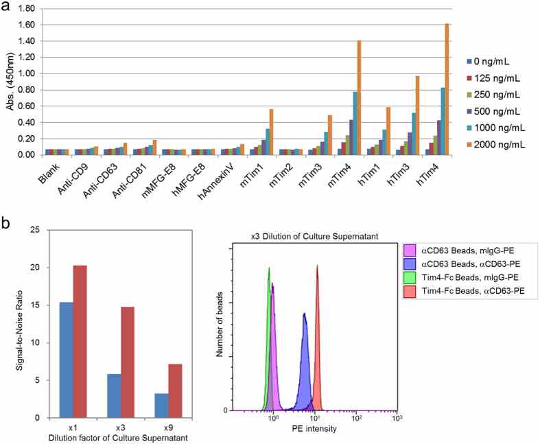
( a ) sEVs in 10K sup of K562 cells (serum free) were purified by ultracentrifugation and the concentration of total proteins was determined by BCA protein assay. The sEVs were serially diluted and then incubated in each well of a Nunc MaxiSorp 96 well plate that had been pre-coated with one of the following capture reagents: anti-CD9, anti-CD63, anti-CD81 antibody, mouse MFG-E8-His, human MFG-E8-His, human
Not For Human Consumption!
Inquiry
- Reviews (0)
- Q&As (0)


Ask a Question for All ANXA8L2 Products
Required fields are marked with *
My Review for All ANXA8L2 Products
Required fields are marked with *