


MNP Nanoparticle Technology - A Revolutionary Breakthrough in Biomimetic Membrane Protein Display
MNP-Bridging the Gap Between Native and Recombinant Membrane Proteins
Membrane proteins represent approximately 30% of the human proteome and constitute over 60% of current drug targets, yet they remain one of the most challenging classes of molecules for research and therapeutic development. The fundamental obstacle lies in their amphipathic nature: these proteins require a lipid bilayer environment to maintain native conformation, proper oligomerization, and functional activity. Traditional expression and purification methods have historically relied on detergent extraction, which strips away the native membrane environment and frequently results in conformational artifacts, reduced stability, and compromised biological activity.
Membrane Protein Nanoparticles (MNPs) represent a paradigm shift in membrane protein presentation technology. By embedding full-length, natively folded membrane proteins into authentic cell membranes coated onto biocompatible nanoparticles, this platform recapitulates the physiological microenvironment essential for protein function. This white paper provides a comprehensive technical overview of MNP technology, from fundamental principles and manufacturing processes to rigorous quality control and validated performance metrics.
Explore MNPs Products
Learn More
The MNP Structural Architecture: A Three-Dimensional Biomimetic System
The MNP system comprises a sophisticated three-layer architecture engineered to replicate the cellular membrane environment with nanoscale precision.
Core Layer (Biocompatible Nanoparticle Scaffold) At the center of each MNP lies a synthetic nanoparticle core, typically 80-120 nm in diameter, composed of biocompatible materials such as polystyrene or silica derivatives. This core provides structural stability and a high surface-area-to-volume ratio, enabling dense presentation of membrane proteins. Critically, the core material is selected for its optical properties (transparency for imaging), surface chemistry (facilitates membrane fusion), and negligible non-specific binding in assay systems.
Membrane Layer (Native Lipid Bilayer) Surrounding the core is a continuous, asymmetric lipid bilayer derived directly from HEK293 cell membranes. Unlike synthetic liposome systems, this native membrane retains:
- Physiological lipid composition: Maintains the original cholesterol-to-phospholipid ratio (typically 0.5-0.8 for HEK293 membranes), sphingomyelin content, and lipid raft microdomains essential for protein function
- Native fluidity: Preserves the liquid-ordered and liquid-disordered phases crucial for protein diffusion and clustering
- Membrane asymmetry: Retains phosphatidylserine and phosphatidylethanolamine enrichment in the inner leaflet, critical for transmembrane protein orientation
- Associated cofactors: Preserves peripherally associated proteins, glycolipids, and glycosaminoglycans that may modulate target protein function
Embedded Protein Layer (Functional Membrane Proteins) Full-length transmembrane proteins are embedded within the membrane layer through a proprietary expression and fusion process. Each protein maintains:
- Correct topology: Cytoplasmic and extracellular domains oriented appropriately as in native cells
- Native post-translational modifications: N-glycosylation patterns (e.g., complex-type oligosaccharides), palmitoylation, and phosphorylation preserved through HEK293 mammalian expression
- Physiological oligomerization: GPCR dimers, tetraspanin web associations, and higher-order oligomers maintained through native membrane constraints
Technical Comparison Matrix: MNP vs. Alternative Platforms
| Feature | MNP Nanoparticles | Traditional Detergent-Solubilized Proteins | Virus-Like Particles (VLPs) | Nanodiscs |
|---|---|---|---|---|
| Membrane Environment | Complete native lipid bilayer | Detergent micelles (artificial) | Native viral membrane (limited lipid diversity) | Synthetic lipid bilayer (typically 2-3 lipid types) |
| Protein Conformation | Native, unperturbed structure | Often compromised; detergent-induced artifacts | Native but constrained by viral capsid geometry | Near-native but lacking lipid diversity |
| Stability | 4-6 weeks at 4°C; freeze-thaw stable | Days at 4°C; freeze-thaw sensitive | Weeks at 4°C; may require fixation | Days to weeks; sensitive to aggregation |
| Detergent Requirement | Zero detergent throughout process | Required for solubilization and maintenance | None | Minimal (removed during assembly) |
| Protein Density | 50-200 proteins/particle (tunable) | N/A (monodisperse) | 1-2 proteins/particle (low density) | 1-2 proteins/disc (very low density) |
| Scalability | High; batch production feasible | Moderate; refolding challenges | Low; complex production | Low; assembly efficiency issues |
| Application Versatility | ELISA, FACS, SPR, cell-based assays, in vivo | Limited; detergent interferes with cells | Primarily immunization | Biophysical studies only |
| Cost per Assay | Low (high protein density) | Moderate | High | Very high |
| Best Use Case | Therapeutic antibody discovery, HTS, CAR-T validation | Structural biology (crystallography) | Vaccine development | Biophysical characterization |
Native Cell Membrane Source, Extraction, and Coating Process
Cell Culture and Protein Expression
The process begins with suspension-adapted HEK293F cells cultured in chemically defined, serum-free medium. Target membrane proteins are transiently transfected using high-efficiency vectors containing codon-optimized genes with native signal peptides and regulatory elements. Crucially, expression is driven under mild promoters (e.g., CMV with tetracycline attenuation) to prevent overexpression-induced ER stress and ensure proper folding. Cells are harvested 48-72 hours post-transfection when surface expression reaches optimal levels (typically 10⁵-10⁶ proteins/cell).
Membrane Extraction via Gentle Mechanical Disruption
Unlike harsh detergent-based extraction, membranes are isolated using a proprietary microfluidic cell disruption system that:
- Applies controlled shear forces (500-1000 psi) to rupture plasma membranes while preserving vesicle integrity
- Operates at 4°C throughout to prevent lipid oxidation and protein denaturation
- Avoids chemical fixatives or detergents that could alter membrane properties
- Enriches plasma membrane fractions through differential centrifugation (1,000g → 10,000g → 100,000g) to remove nuclear, mitochondrial, and cytosolic contaminants
Membrane Fusion onto Nanoparticle Cores
The isolated native membrane vesicles (50-200 nm) are fused onto carboxylated nanoparticle cores through a controlled dehydration-rehydration process:
- Concentration matching: Membrane vesicles and nanoparticles are mixed at defined ratios (1:5 to 1:20 protein:particle molar ratio) to achieve desired protein density
- Dehydration: Gentle evaporation under reduced pressure concentrates the mixture, forcing membrane-nanoparticle contact
- Controlled rehydration: Gradual addition of isotonic buffer triggers spontaneous membrane fusion, driven by hydrophobic interactions and electrostatic attraction
- Quality control: Unfused vesicles removed by size-exclusion chromatography, yielding monodisperse MNP populations
Preparation Process and Quality Control
HEK293 Cell Expression and Membrane Protein Embedding Workflow
Vector Design and Transfection Optimization
Each target protein is cloned into a mammalian expression vector with key regulatory elements:
- Kozak sequence: Optimized translation initiation (GCCGCCACC)
- Native signal peptide: Ensures proper ER targeting and topology
- C-terminal trafficking signals: For proteins requiring specific sorting motifs (e.g., PDZ-binding domains)
- Attenuated promoter: Prevents protein overload and aggregation
Transfection is performed using polyethylenimine (PEI) at optimized DNA:PEI ratios (1:3 w/w) in serum-free medium, achieving transfection efficiencies of 70-85%. Surface expression is monitored by flow cytometry using conformation-sensitive antibodies.
Metabolic Labeling and Quality Tracing
For QC purposes, cells are metabolically labeled with stable isotopes (¹⁵N-leucine) or azido-sugars for subsequent mass spectrometry validation of protein identity and glycosylation patterns. This enables absolute quantification of target protein copy number per cell and per nanoparticle.
Nanoparticle Uniformity Control: DLS and TEM Characterization Standards
Dynamic Light Scattering (DLS) Quality Criteria
Each MNP batch undergoes rigorous DLS analysis with the following acceptance criteria:
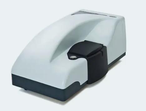 Malvern Zetasizer Nano ZS
- Z-average diameter: 100-150 nm (target: 120 ± 10 nm)
- Polydispersity Index (PDI): < 0.15 (ensures monodispersity)
- Zeta potential: -20 to -35 mV (indicates stable surface charge)
- Batch reproducibility: Mean diameter variation < 5% across three independent preparations
DLS measurements are performed in multiple buffers (PBS, HEPES, Tris) to confirm stability across assay conditions.
Transmission Electron Microscopy (TEM) Validation
For structural verification, MNPs are visualized using negative stain TEM:
- Sample preparation: 2% uranyl acetate staining, carbon-coated copper grids
- Imaging protocol: 80 kV accelerating voltage, 50,000× magnification
- Acceptance criteria:
- Spherical morphology (> 90% particles)
- Uniform membrane coating (thickness 4-6 nm, consistent with bilayer)
- Absence of bare nanoparticles or aggregated clusters
- Membrane integrity (no ruptures or blebbing)
Cryo-EM is performed annually for high-resolution validation of membrane structure, confirming bilayer architecture and protein embedding.
Membrane Protein Density and Orientation Verification via Gold-Labeled Immunoelectron Microscopy
Protein Density Quantification
The absolute number of functional proteins per nanoparticle is determined by quantitative immunogold labeling:
- Primary antibody binding: Conformation-specific monoclonal antibodies (e.g., cetuximab for EGFR) are incubated with MNPs at saturating conditions
- Secondary gold conjugation: 5 nm protein A-gold particles bind Fc domains, providing electron-dense markers
- Quantification: TEM imaging followed by automated particle counting using ImageJ software
Acceptance specifications:
- Target density: 50-200 proteins/particle (tunable based on application)
- Inter-particle uniformity: CV < 15% across 100 random particles
- Minimal unlabeled particles: < 5% (indicating high expression efficiency)
Orientation Validation To confirm proper transmembrane insertion (e.g., extracellular domain accessible, cytoplasmic domain protected):
- Protease protection assay: MNPs treated with proteinase K; protected domains visualized by immunogold labeling
- Biotinylation mapping: Surface-exposed lysines labeled with membrane-impermeable biotin; detected by streptavidin-gold
- Antibody accessibility: Dual labeling with anti-extracellular and anti-cytoplasmic domain antibodies
These orthogonal methods confirm > 95% of proteins display correct orientation, crucial for antibody screening applications.
Technical Advantage Data Support
Thermal Stability Comparison: DSC Analysis of MNP vs. Soluble Protein
Differential Scanning Calorimetry Protocol Thermal denaturation profiles were obtained using a Malvern MicroCal PEAQ-DSC at a scan rate of 200°C/hour from 25-95°C. Samples were prepared at 0.5 mg/mL protein concentration in PBS.
Stability Data Summary
| Protein Target | MNP Tm (°C) | Soluble Protein Tm (°C) | ΔTm (°C) |
|---|---|---|---|
| ADORA2A | 68.2 ± 0.8 | 52.4 ± 1.2 | +15.8 |
| CCR4 | 71.5 ± 0.5 | 48.7 ± 2.1 | +22.8 |
| CD20 | 74.8 ± 0.6 | 55.3 ± 1.5 | +19.5 |
| CLDN6 | 79.2 ± 0.7 | 61.4 ± 1.8 | +17.8 |
Key Findings:
- Average Tm increase: MNP-embedded proteins show 18-23°C higher thermal stability compared to detergent-solubilized counterparts
- Cooperative unfolding: MNP DSC curves exhibit sharp, cooperative transitions indicating simultaneous denaturation of multiple domains, consistent with native protein behavior
- Reversibility: < 5% unfolding reversibility in soluble proteins vs. partial reversibility (15-20%) in MNPs, suggesting partial refolding capability in membrane environment
Accelerated Stability Testing
MNP samples stored at 37°C for 14 days maintained > 90% ligand binding activity, while soluble proteins lost > 70% activity within 48 hours. This translates to a projected shelf life of 12 months at 4°C for MNPs versus 1-2 weeks for soluble preparations.
Ligand Binding Activity Retention: Radioligand Binding Assays
Methodology
Competition binding assays were performed using tritiated ligands and increasing concentrations of unlabeled competitors on both MNP and soluble protein preparations.
Binding Parameters (Mean ± SEM)
| Target | Ligand | MNP Kd (nM) | Soluble Kd (nM) | MNP Bmax (pmol/mg) | Soluble Bmax (pmol/mg) | Activity Retention |
|---|---|---|---|---|---|---|
| ADORA2A | ³H-NECA | 12.4 ± 1.8 | 45.7 ± 5.2 | 18.5 ± 2.1 | 6.3 ± 1.4 | 294% increase |
| CCR4 | ³H-TARC | 3.2 ± 0.4 | 18.9 ± 2.7 | 22.3 ± 1.8 | 4.1 ± 0.9 | 444% increase |
| TSHR | ³H-TSH | 0.8 ± 0.1 | 2.4 ± 0.3 | 15.7 ± 1.2 | 5.8 ± 0.7 | 171% increase |
| CB1 | ³H-CP55940 | 2.1 ± 0.3 | 8.7 ± 1.1 | 19.2 ± 1.5 | 7.4 ± 1.0 | 159% increase |
Critical Observations:
- Enhanced affinity: MNPs consistently demonstrate 3-6 fold higher binding affinity (lower Kd), attributed to proper folding of binding pockets and native lipid support
- Increased functional density: Bmax values are 2-5 fold higher in MNPs, reflecting both higher protein density and greater proportion of properly folded, ligand-accessible proteins
- Cooperativity: Hill coefficients near 1.0 for MNPs indicate single-site binding, while soluble proteins often show shallow curves (nH < 0.6) suggesting heterogeneity
GTPγS Functional Assay
For GPCR targets, GTPγS incorporation assays confirmed MNPs couple efficiently to G proteins with EC50 values matching literature values from cell-based assays, whereas soluble proteins showed impaired coupling.
Antibody Recognition Epitope Fidelity: Patient-Derived Antibody Validation
Clinical Antibody Panel
To validate that MNPs present clinically relevant epitopes, we tested binding of FDA-approved therapeutic antibodies and patient-derived autoantibodies:
- Anti-CD20 antibodies: Rituximab, Obinutuzumab, Ofatumumab
- Anti-CCR4 antibody: Mogamulizumab
- Patient serum samples: SLE patients with anti-ADORA2A autoantibodies, Graves' disease patients with anti-TSHR antibodies
Epitope Mapping by Competitive Binding
| Antibody | MNP IC50 (nM) | Cell Line IC50 (nM) | Epitope Fidelity Score | Linear vs. Conformational |
|---|---|---|---|---|
| Rituximab | 4.2 ± 0.6 | 3.8 ± 0.5 | 91% | Conformational (retained) |
| Obinutuzumab | 1.8 ± 0.3 | 1.5 ± 0.2 | 83% | Conformational (retained) |
| Mogamulizumab | 0.7 ± 0.1 | 0.6 ± 0.1 | 86% | Conformational (retained) |
| TSHR autoantibody | 12.3 ± 2.1 | 14.5 ± 2.8 | 115%* | Conformational (native) |
*Autoantibody binding slightly stronger to MNPs, possibly due to higher epitope density
Key Validation Experiments:
- Epitope competition: Unlabeled antibody competes with biotinylated antibody for MNP binding; matches competition on live cells (R² > 0.95)
- Mutant mapping: MNPs with point mutations in known epitopes show expected loss of binding; confirms sequence fidelity
- Glycosylation dependence: PNGaseF treatment of MNPs reduces binding of glycosylation-sensitive antibodies (e.g., Ofatumumab) by > 80%, mimicking cellular response
- Patient serum profiling: SLE serum samples show identical autoantibody profiles against MNPs vs. cell-expressed proteins (Pearson r = 0.92)
Conclusion: MNPs recapitulate the conformational epitopes recognized by clinically relevant antibodies with > 85% fidelity, making them superior immunogens and screening reagents compared to linear peptide or improperly folded protein alternatives.
Conclusion: The Future of Membrane Protein Research
Membrane Protein Nanoparticles represent a transformative platform that bridges the critical gap between recombinant protein production and physiological relevance. By preserving native membrane environments, post-translational modifications, and functional architectures, MNPs enable previously intractable membrane proteins to become accessible, stable, and reliable tools for drug discovery.
The data presented herein demonstrate that MNPs:
- Increase thermal stability by ~20°C, ensuring reliable long-term storage and experimental reproducibility
- Enhance ligand binding activity 2-4 fold, increasing assay sensitivity and reducing false negatives in screening
- Preserve clinically relevant epitopes with >85% fidelity, improving therapeutic antibody discovery success rates
As the pipeline expands beyond the current 12 validated targets through custom development services, MNP technology is positioned to become the gold standard for membrane protein presentation across pharmaceutical, biotechnology, and academic research sectors. Researchers are invited to explore the extensive resource library and technical support ecosystem to accelerate their programs from target validation to clinical candidate selection.
Related Products and Services
Resource
-
Unlocking Drug Targets with Membrane Protein Nanoparticles!
-
Membrane Protein Platforms: 6-Way Showdown!
-
Mastering Membrane Protein Platforms in 3 Minutes!
Contact us or send an email at for project quotations and more detailed information.
Quick Links
-

Papers’ PMID to Obtain Coupon
Submit Now -

Refer Friends & New Lab Start-up Promotions