


Recombinant Human Cadherin 2, Type 1, N-cadherin (Neuronal)
| Cat.No. : | CDH2-87H |
| Product Overview : | Recombinant Human Cadherin-N protein 741-907 a.a. expressed inE.coli,43 kDa. |
- Specification
- Gene Information
- Related Products
- Citation
- Download
| Species : | Human |
| Source : | E.coli |
| Tag : | Non |
| Protein Length : | 741-907 a.a. |
| Description : | Cadherins comprise a family of Ca+ dependent adhesion molecules that function to mediate cell-cell binding critical to the maintenance of tissue structure and morphogenesis. The classical cadherins, E-, N- and P-cadherin, consist of large extracellular domains characterized by a series of five homologous NH2 terminal repeats. The most distal of these cadherins is thought to be responsible for binding specificity, transmembrane domains and carboxy terminal intracellular domains. |
| Presentation : | Recombinant Human Cadherin-N protein at 100µg/ml in 50mM Tris-Acetate, pH7.5, 1mM EDTA and 20% Glycerol. |
| Characterization : | On SDS-PAGE commassie blue stained gel, the purified recombinant protein shows a band at 43 kDa. |
| Applications : | •ELISA •Inhibition Assays •Western Blotting |
| Storage : | Store vial at -20°Cto -80°C. When stored at the recommended temperature, this protein is stable for 12 months. |
| Gene Name | CDH2 cadherin 2, type 1, N-cadherin (neuronal) [ Homo sapiens ] |
| Synonyms | CDH2; cadherin 2, type 1, N-cadherin (neuronal); CDHN; NCAD; CD325; CDw325; cadherin 2, type 1; N-cadherin 1; neural cadherin; cadherin 2, N-cadherin (neuronal); calcium-dependent adhesion protein, neuronal; 5` partial; N-cadherin; Cadherin-2;CD325 antigen |
| Gene ID | 1000 |
| mRNA Refseq | NM_001792 |
| Protein Refseq | NP_001783 |
| MIM | 114020 |
| UniProt ID | P19022 |
| Chromosome Location | 18q12.1 |
| Pathway | Arrhythmogenic right ventricular cardiomyopathy (ARVC); Cell adhesion molecules (CAMs); Cell-cell adhesion systems |
| Function | RPTP-like protein binding; beta-catenin binding; calcium ion binding; protein kinase binding; protein phosphatase binding |
| ◆ Recombinant Proteins | ||
| CDH2-7023H | Recombinant Human CDH2 protein, His-tagged | +Inquiry |
| RFL25845GF | Recombinant Full Length Chicken Cadherin-2(Cdh2) Protein, His-Tagged | +Inquiry |
| CDH2-1742R | Recombinant Rhesus Monkey CDH2 Protein, hIgG4-tagged | +Inquiry |
| Cdh2-7024M | Recombinant Mouse Cdh2 protein, His-tagged | +Inquiry |
| CDH2-948R | Recombinant Rat CDH2 Protein, His (Fc)-Avi-tagged | +Inquiry |
| ◆ Cell & Tissue Lysates | ||
| CDH2-971MCL | Recombinant Mouse CDH2 cell lysate | +Inquiry |
| CDH2-980HCL | Recombinant Human CDH2 cell lysate | +Inquiry |
Rickettsia Sca4 reduces vinculin-mediated intercellular tension to promote spread
Journal: Cell PubMed ID: 27768890 Data: 2017/10/20
Authors: Rebecca L. Lamason, Effie Bastounis, Matthew D. Welch
Article Snippet:To measure traction forces mediated by E-cadherin, activated gels were first coated with 0.2 mg/ml rabbit anti-human IgG (Fcγ-fragment specific; Jackson ImmunoResearch) and incubated overnight at 4°C.To measure traction forces mediated by E-cadherin, activated gels were first coated with 0.2 mg/ml rabbit anti-human IgG (Fcγ-fragment specific; Jackson ImmunoResearch) and incubated overnight at 4°C.. Gels were washed with PBS the next day to remove unbound antibody and incubated for 3 h at 4°C with 50 μg/ml recombinant human E-cadherin/Fc chimera (Creative BioMart).. Gels were then washed with PBS, blocked for 30 min at room temperature with 1% BSA/PBS, washed again with PBS, and equilibrated in DMEM + 10% FBS for 30 min at 37°C prior to adding cells.Gels were then washed with PBS, blocked for 30 min at room temperature with 1% BSA/PBS, washed again with PBS, and equilibrated in DMEM + 10% FBS for 30 min at 37°C prior to adding cells.
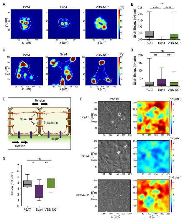
TFM results for individual cells adherent to polyacrylamide gels coated with
Not For Human Consumption!
Inquiry
- Reviews (0)
- Q&As (0)


Ask a Question for All CDH2 Products
Required fields are marked with *
My Review for All CDH2 Products
Required fields are marked with *