


Recombinant Human OLFM4 protein, His-tagged
| Cat.No. : | OLFM4-3222H |
| Product Overview : | Recombinant Human OLFM4 protein(NP_006409.3) (Met 1-Gln 510) was expressed in HEK293, fused with a polyhistidine tag at the C-terminus. |
| Availability | July 28, 2026 |
| Unit | |
| Price | |
| Qty |
- Specification
- Gene Information
- Related Products
- Citation
- Download
| Species : | Human |
| Source : | HEK293 |
| Tag : | His |
| Protein Length : | 1-510 aa |
| Form : | Lyophilized from sterile PBS, pH 7.4. |
| Molecular Mass : | The recombinant human OLFM4 consists of 501 amino acids and predictes a molecular mass of 56.6 kDa. In SDS-PAGE under reducing conditions, the apparent molecular mass of rh OLFM4 is approximately 65-70 kDa due to different glycosylation. |
| Endotoxin : | < 1.0 EU per μg of the protein as determined by the LAL method. |
| Purity : | > 92 % as determined by SDS-PAGE |
| Storage : | Store it under sterile conditions at -20°C to -80°C upon receiving. Recommend to aliquot the protein into smaller quantities for optimal storage. Avoid repeated freeze-thaw cycles. |
| Reconstitution : | It is recommended that sterile water be added to the vial to prepare a stock solution of 0.2 ug/ul. Centrifuge the vial at 4°C before opening to recover the entire contents. |
| Gene Name | OLFM4 olfactomedin 4 [ Homo sapiens ] |
| Official Symbol | OLFM4 |
| Synonyms | OLFM4; olfactomedin 4; olfactomedin-4; GC1; GW112; OlfD; antiapoptotic protein GW112; G-CSF-stimulated clone 1 protein; OLM4; hGC-1; hOLfD; UNQ362; bA209J19.1; KIAA4294; |
| Gene ID | 10562 |
| mRNA Refseq | NM_006418 |
| Protein Refseq | NP_006409 |
| MIM | 614061 |
| UniProt ID | Q6UX06 |
| ◆ Recombinant Proteins | ||
| OLFM4-3222H | Recombinant Human OLFM4 protein, His-tagged | +Inquiry |
| OLFM4-3222M | Recombinant Mouse OLFM4 Protein | +Inquiry |
| OLFM4-3434H | Recombinant Human OLFM4 Protein (Ile407-Gln510), His tagged | +Inquiry |
| Olfm4-136M | Recombinant Mouse Olfm4 Protein, His-tagged | +Inquiry |
| Olfm4-9170M | Recombinant Mouse Olfm4, His-tagged | +Inquiry |
| ◆ Cell & Tissue Lysates | ||
| OLFM4-2429HCL | Recombinant Human OLFM4 cell lysate | +Inquiry |
| OLFM4-384HKCL | Human OLFM4 Knockdown Cell Lysate | +Inquiry |
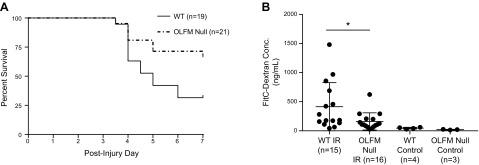
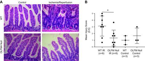
The olfactomedin-4 positive neutrophil has a role in murine intestinal ischemia/reperfusion injury
Journal: The FASEB Journal PubMed ID: 31593636 Data: 2020/11/25
Authors: Nick C. Levinsky, Jaya Mallela, Matthew N. Alder
Article Snippet:To evaluate whether secreted OLFM4 may have an effect on surrounding inflammatory cells, murine RAW264 cells were grown in tissue culture with DMEM with 10% fetal bovine serum and plated in 6-well plates at 1 × 10 6 cells/well until reaching 50–60% confluency.To evaluate whether secreted OLFM4 may have an effect on surrounding inflammatory cells, murine RAW264 cells were grown in tissue culture with DMEM with 10% fetal bovine serum and plated in 6-well plates at 1 × 10 6 cells/well until reaching 50–60% confluency.. They were then stimulated overnight with 10–100 ng/ml of recombinant murine OLFM4 (Creative BioMart, Shirley, NY, USA) at 37°C.. Control treatments included medium alone or 1 μg/ml of LPS.Control treatments included medium alone or 1 μg/ml of LPS.
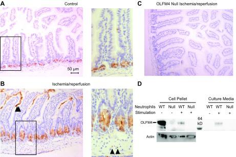
Representative immunohistochemical sections stained for OLM4 in mouse intestine. A) In healthy control WT mice,
Not For Human Consumption!
Inquiry
- Reviews (0)
- Q&As (0)


Ask a Question for All OLFM4 Products
Required fields are marked with *
My Review for All OLFM4 Products
Required fields are marked with *