


Recombinant Human TLR9 protein
| Cat.No. : | TLR9-452H |
| Product Overview : | Recombinant Human TLR9 was expressed in Wheat Germ. |
- Specification
- Gene Information
- Related Products
- Citation
- Download
| Species : | Human |
| Source : | Wheat Germ |
| Tag : | Non |
| Description : | The protein encoded by this gene is a member of the Toll-like receptor (TLR) family which plays a fundamental role in pathogen recognition and activation of innate immunity. TLRs are highly conserved from Drosophila to humans and share structural andfunctional similarities. They recognize pathogen-associated molecular patterns (PAMPs) that are expressed on infectious agents, and mediate the production of cytokines necessary for the development of effective immunity. The various TLRs exhibit different patterns of expression. This gene is preferentially expressed in immune cell rich tissues, such as spleen, lymph node, bone marrow and peripheral blood leukocytes. Studies in mice and human indicate that this receptor mediates cellular response to unmethylatedCpG dinucleotides in bacterial DNA to mount an innate immune response. |
| Form : | 25 mM Tris-HCl of pH8.0 containing 2% glycerol. |
| Molecular Mass : | 113.52 kDa |
| AA Sequence : | MGFCRSALHPLSLLVQAIMLAMTLALGTLPAFLPCELQPHGLVNCNWLFLKSVPHFSMAAPRGNVTSLSLSSNRI HHLHDSDFAHLPSLRHLNLKWNCPPVGLSPMHFPCHMTIEPSTFLAVPTLEELNLSYNNIMTVPALPKSLISLSL SHTNILMLDSASLAGLHALRFLFMDGNCYYKNPCRQALEVAPGALLGLGNLTHLSLKYNNLTVVPRNLPSSLEYL LLSYNRIVKLAPEDLANLTALRVLDVGGNCRRCDHAPNPCMECPRHFPQLHPDTFSHLSRLEGLVLKDSSLSWLN ASWFRGLGNLRVLDLSENFLYKCITKTKAFQGLTQLRKLNLSFNYQKRVSFAHLSLAPSFGSLVALKELDMHGIF FRSLDETTLRPLARLPMLQTLRLQMNFINQAQLGIFRAFPGLRYVDLSDNRISGASELTATMGEADGGEKVWLQP GDLAPAPVDTPSSEDFRPNCSTLNFTLDLSRNNLVTVQPEMFAQLSHLQCLRLSHNCISQAVNGSQFLPLTGLQV LDLSHNKLDLYHEHSFTELPRLEALDLSYNSQPFGMQGVGHNFSFVAHLRTLRHLSLAHNNIHSQVSQQLCSTSL RALDFSGNALGHMWAEGDLYLHFFQGLSGLIWLDLSQNRLHTLLPQTLRNLPKSLQVLRLRDNYLAFFKWWSLHF LPKLEVLDLAGNQLKALTNGSLPAGTRLRRLDVSCNSISFVAPGFFSKAKELRELNLSANALKTVDHSWFGPLAS ALQILDVSANPLHCACGAAFMDFLLEVQAAVPGLPSRVKCGSPGQLQGLSIFAQDLRLCLDEALSWDCFALSLLA VALGLGVPMLHHLCGWDLWYCFHLCLAWLPWRGRQSGRDEDALPYDAFVVFDKTQSAVADWVYNELRGQLEECRG RWALRLCLEERDWLPGKTLFENLWASVYGSRKTLFVLAHTDRVSGLLRASFLLAQQRLLEDRKDVVVLVILSPDG RRSRYVRLRQRLCRQSVLLWPHQPSGQRSFWAQLGMALTRDNHHFYNRNFCQGPTAE |
| Applications : | Antibody Production;Functional Study: Recommended usage only, not validated yet;Compound Screening: Recommended usage only, not validated yet. |
| Notes : | Heating may cause protein aggregation. Please do not heat this product before electrophoresis. Best use within three months from the date of receipt of this protein. |
| Storage : | Store at -80 centigrade. Aliquot to avoid repeated freezing and thawing. |
| Gene Name | TLR9 toll-like receptor 9 [ Homo sapiens ] |
| Official Symbol | TLR9 |
| Synonyms | TLR9; toll-like receptor 9; CD289; |
| Gene ID | 54106 |
| mRNA Refseq | NM_017442 |
| Protein Refseq | NP_059138 |
| MIM | 605474 |
| UniProt ID | Q9NR96 |
| Chromosome Location | 3p21.3 |
| Pathway | African trypanosomiasis, organism-specific biosystem; African trypanosomiasis, conserved biosystem; Chagas disease (American trypanosomiasis), organism-specific biosystem; Chagas disease (American trypanosomiasis), conserved biosystem; Herpes simplex infection, organism-specific biosystem; Herpes simplex infection, conserved biosystem; IRS-mediated signalling, organism-specific biosystem; |
| Function | interleukin-1 receptor binding; receptor activity; siRNA binding; |
| ◆ Recombinant Proteins | ||
| Tlr9-697R | Recombinant Rat Tlr9 protein, His & T7-tagged | +Inquiry |
| TLR9-1684R | Recombinant Rhesus Monkey TLR9 Protein, hIgG4-tagged | +Inquiry |
| TLR9-452H | Recombinant Human TLR9 protein | +Inquiry |
| TLR9-695H | Recombinant Human TLR9 protein, His-tagged | +Inquiry |
| TLR9-422R | Recombinant Rabbit TLR9 Protein, His-tagged | +Inquiry |
| ◆ Cell & Tissue Lysates | ||
| TLR9-1042HCL | Recombinant Human TLR9 293 Cell Lysate | +Inquiry |
Toll-Like Receptor 9-Mediated Neuronal Innate Immune Reaction Is Associated with Initiating a Pro-Regenerative State in Neurons of the Dorsal Root Ganglia Non-Associated with Sciatic Nerve Lesion
Journal: International Journal of Molecular Sciences PubMed ID: 34299065 Data: 2021/7/12
Authors: Petr Dubovy, Ivana Hradilová-Sví?enská, Ilaria Tonazzini
Article Snippet:Another portion was incubated with mouse monoclonal anti-TLR9 (1:100; sc-52966, Santa Cruz Biotechnology, Heidelberg, Germany) and one of either rabbit monoclonal anti–Rab7 (1:100; #9367, Cell Signaling Technology, The Netherlands) or rabbit polyclonal anti-GRP78 (1:500; ab53068, Abcam, Cambridge, UK) antibodies.one of either rabbit monoclonal anti–Rab7 (1:100; #9367, Cell Signaling Technology, The Netherlands) or rabbit polyclonal anti-GRP78 (1:500; ab53068, Abcam, Cambridge, UK) antibodies. ... A mixture (1:1) of affinity purified TRITC-conjugated donkey anti-rabbit and FITC-conjugated donkey anti-mouse secondary antibodies (1:100; AP182R and AP192F, EMD Millipore, Tamecula, CA, USA) was applied at room temperature for 90 min. Control sections were incubated without the primary antibodies, with preabsorbed polyclonal TLR9 antibody (20 μg/mL, TLR9 blocking peptide, MBS-152758, MyBioSource, San Diego, CA, USA), preabsorbed monoclonal antibody using recombinant human TLR9 protein (10 μg/mL, TLR9-48H, Creative-Biomart, Shirley, NY, USA) or with a reverse combination of primary and secondary antibodies.. The control sections displayed no immunostaining.The control sections displayed no immunostaining.
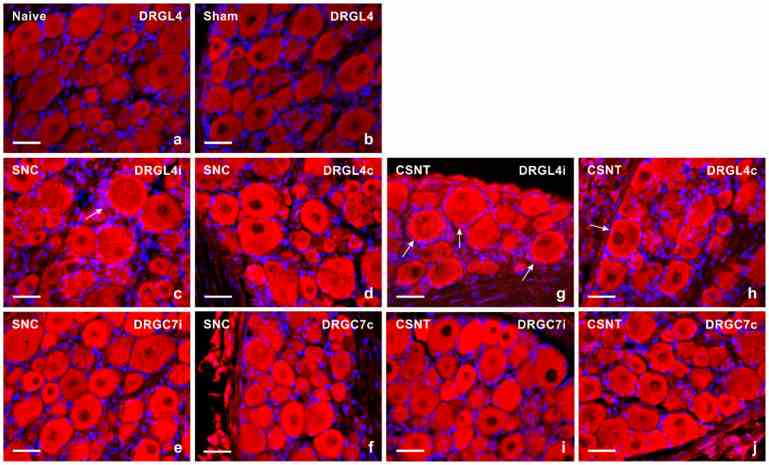
Representative sections of DRG from naive rat ( a ) and rats with sterile sham- ( b ), SNC- ( c – f ) or CSNT- ( g – j ) interventions for 7 days. The DRG sections of lumbar (DRG-L4) and cervical (DRG-C7) segments of both ipsilateral ( i ) and contralateral ( c ) sides were immunostained for

Results from image analysis of
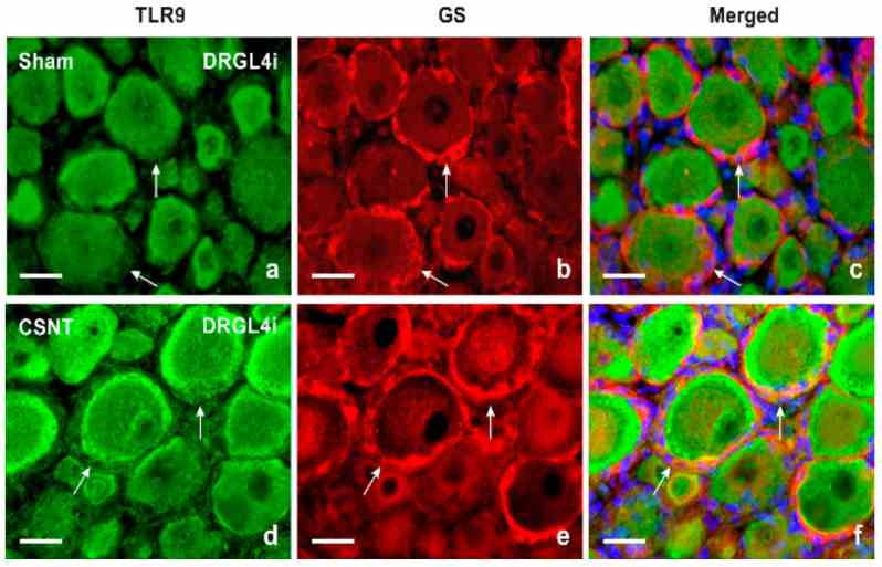
Double immunofluorescence staining for
Not For Human Consumption!
Inquiry
- Reviews (0)
- Q&As (0)


Ask a Question for All TLR9 Products
Required fields are marked with *
My Review for All TLR9 Products
Required fields are marked with *