


Recombinant Full Length Human KRT19 Protein, His-tagged 
| Cat.No. : | KRT19-7239H |
| Product Overview : | Recombinant Full Length Human KRT19 Protein with His tag was expressed in E. coli. |
| Availability | July 28, 2026 |
| Unit | |
| Price | |
| Qty |
- Specification
- Gene Information
- Related Products
- Citation
- Download
| Species : | Human |
| Source : | E.coli |
| Tag : | His |
| Protein Length : | 1-400 aa |
| Description : | The protein encoded by this gene is a member of the keratin family. The keratins are intermediate filament proteins responsible for the structural integrity of epithelial cells and are subdivided into cytokeratins and hair keratins. The type I cytokeratins consist of acidic proteins which are arranged in pairs of heterotypic keratin chains. Unlike its related family members, this smallest known acidic cytokeratin is not paired with a basic cytokeratin in epithelial cells. It is specifically expressed in the periderm, the transiently superficial layer that envelopes the developing epidermis. The type I cytokeratins are clustered in a region of chromosome 17q12-q21. |
| Tag : | His |
| Molecular Mass : | 47 kDa |
| AA Sequence : | MGSSHHHHHHSSGLVPRGSHMGSMTSYSYRQSSATSSFGGLGGGSVRFGPGVAFRAPSIHGGSGGRGVSVSSARFVSSSSSGAYGGGYGGVLTASDGLLAGNEKLTMQNLNDRLASYLDKVRALEAANGELEVKIRDWYQKQGPGPSRDYSHYYTTIQDLRDKILGATIENSRIVLQIDNARLAADDFRTKFETEQALRMSVEADINGLRRVLDELTLARTDLEMQIEGLKEELAYLKKNHEEEISTLRGQVGGQVSVEVDSAPGTDLAKILSDMRSQYEVMAEQNRKDAEAWFTSRTEELNREVAGHTEQLQMSRSEVTDLRRTLQGLEIELQSQLSMKAALEDTLAETEARFGAQLAHIQALISGIEAQLGDVRADSERQNQEYQRLMDIKSRLEQEIATYRSLLEGQEDHYNNLSASKVL |
| Purity : | >90% by SDS-PAGE |
| Storage : | Store it under sterile conditions at -20 to -80 °C. It is recommended that the protein be aliquoted for optimal storage. Avoid repeated freeze-thaw cycles. |
| Concentration : | 1mg/mL by BCA |
| Storage Buffer : | Sterile PBS, pH7.4 |
| Publications : |
| Gene Name | KRT19 keratin 19 [ Homo sapiens (human) ] |
| Official Symbol | KRT19 |
| Synonyms | KRT19; keratin 19; keratin, type I cytoskeletal 19; 40 kDa keratin intermediate filament; CK19; cytokeratin 19; K1CS; K19; keratin; type I cytoskeletal 19; type I; 40 kd; MGC15366; CK-19; keratin-19; cytokeratin-19; keratin, type I, 40-kd; 40-kDa keratin intermediate filament; |
| Gene ID | 3880 |
| mRNA Refseq | NM_002276 |
| Protein Refseq | NP_002267 |
| MIM | 148020 |
| UniProt ID | P08727 |
| ◆ Recombinant Proteins | ||
| KRT19-616H | Recombinant Human Keratin 19 | +Inquiry |
| KRT19-67M | Recombinant Mouse KRT19 Protein, His-tagged | +Inquiry |
| Krt19-1284M | Recombinant Mouse Krt19 Protein, MYC/DDK-tagged | +Inquiry |
| KRT19-4395H | Recombinant Human KRT19 Protein (Glu80-Pro241), N-GST tagged | +Inquiry |
| KRT19-019H | Recombinant Human KRT19, MYC/DDK-tagged | +Inquiry |
| ◆ Native Proteins | ||
| KRT19-169H | Native Human Cytokeratin 19 | +Inquiry |
| KRT19-5H | Native Human CK19 | +Inquiry |
| KRT19-40H | Native Human KRT19 protein | +Inquiry |
| KRT19-382H | Native Human KRT19 | +Inquiry |
| ◆ Cell & Tissue Lysates | ||
| KRT19-4875HCL | Recombinant Human KRT19 293 Cell Lysate | +Inquiry |
Pancreatic cancer cells assemble a CXCL12-keratin 19 coating to resist immunotherapy
Journal: bioRxiv Data: 2020/9/4
Authors: Wang Zhikai, Yan Ran, Fearon Douglas T.
Article Snippet:PrePrint: The buffer of commercially available recombinant human KRT19 protein (Creative BioMart, KRT19-7239H) was changed to protein binding buffer (25 mM Tris, pH 7.5, 100 mM NaCl, 1% Triton X-100), using Zeba spin desalting columns (Thermo, 89882).. For protein binding, biotinylated recombinant human CXCL12 (Chemotactics, B-CXCL12) or CXCL8 (Chemotactics, B-CXCL8) was incubated with KRT19 at 4°C for 2 hours.For protein binding, biotinylated recombinant human CXCL12 (Chemotactics, B-CXCL12) or CXCL8 (Chemotactics, B-CXCL8) was incubated with KRT19 at 4°C for 2 hours.
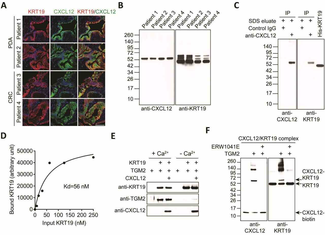
( A ) Sections of freshly resected human PDA and CRC were stained with fluorochrome-conjugated antibodies to
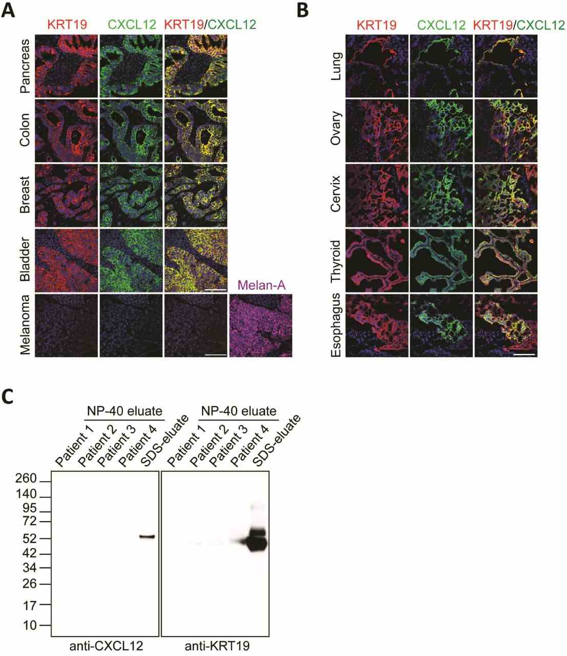
( A - B ) Sections of FFPE human carcinomas and melanoma (A) and frozen human carcinomas (B) were stained with fluorescent antibodies to
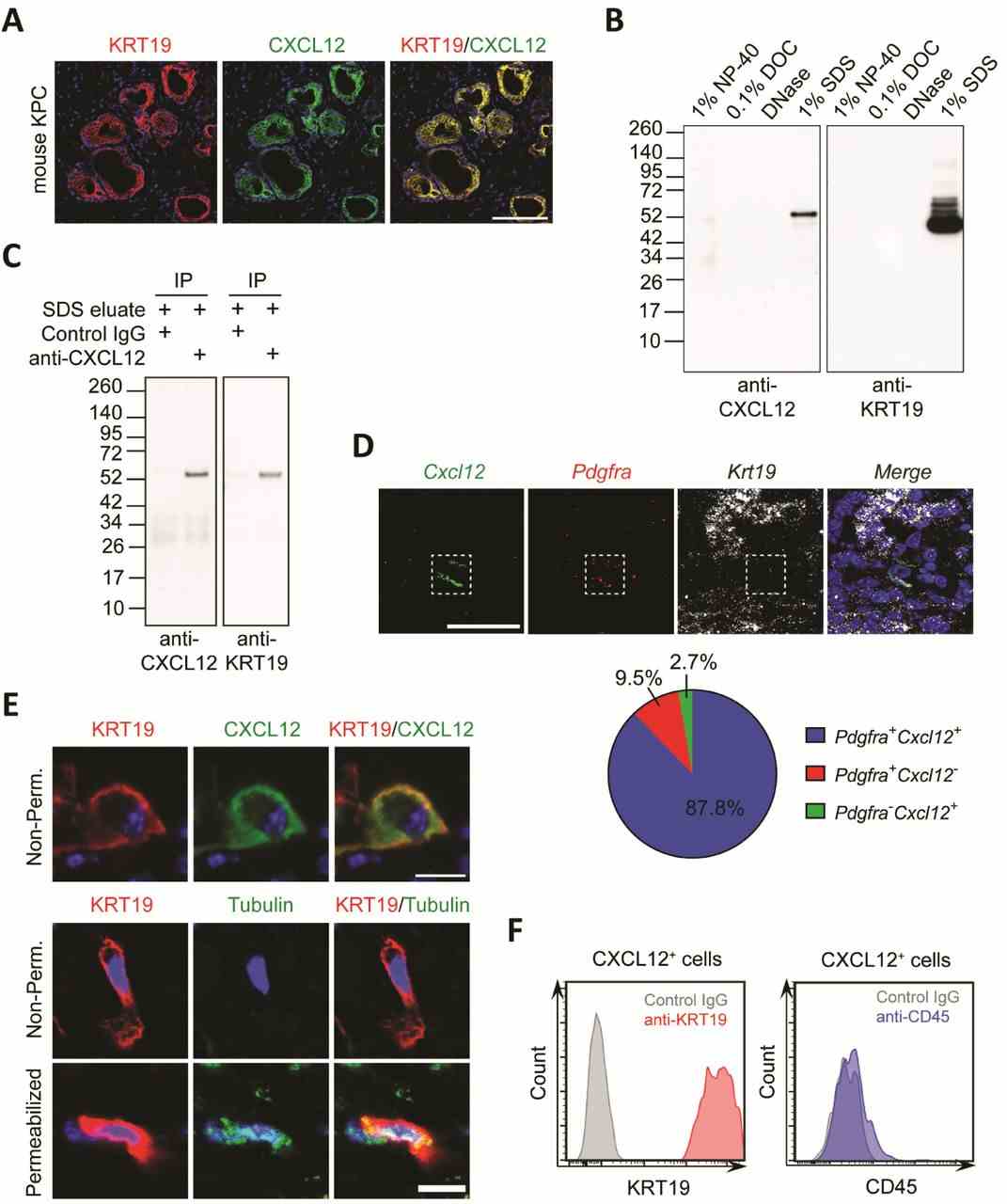
( A ) A frozen section of mouse autochthonous KPC tumor was stained with fluorescent antibodies to
Not For Human Consumption!
Inquiry
- Reviews (0)
- Q&As (0)


Ask a Question for All KRT19 Products
Required fields are marked with *
My Review for All KRT19 Products
Required fields are marked with *