NQO1 regulates mitotic progression and response to mitotic stress through modulating SIRT2 activity
Journal: Free radical biology & medicine PubMed ID: 30114477 Data: 2019/10/1
Authors: Hong-Jun Kang, Ha Yong Song, Athanassios Vassilopoulos
Article Snippet:Recombinant human SIRT2 and GST tagged NQO1 (GST-NQO1) proteins were purchased from Abcam and Creative BioMart, respectively.. SIRT2 and GST-NQO1 were incubated for 1 hour at 4°C with rotation in 20 mM Tris-HCl (pH 7.62), 150 mM NaCl and 1% Triton X-100 binding buffer.SIRT2 and GST-NQO1 were incubated for 1 hour at 4°C with rotation in 20 mM Tris-HCl (pH 7.62), 150 mM NaCl and 1% Triton X-100 binding buffer.
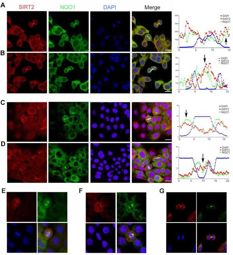
NQO1 and SIRT2 colocalize to the mitotic spindle during mitosis. (A, B) Confocal microscopy images show the locations of endogenous SIRT2 (red) and NQO1 (green) during both metaphase (A) and telophase (B) in MCF-7 human breast cancer cells. (C, D) Confocal microscopy images show the locations of endogenous SIRT2 (red) and NQO1 (green) during both metaphase (C) and telophase (D) in BxPC-3 human pancreatic cancer cells. In all panels above, DNA was stained with DAPI (blue). On the right, graphs represent signal intensity scans along the lines drawn for each panel. Scale bar, 20 μm. (E, F, G) Higher magnification images (60x) under same experimental conditions as shown in A, B.
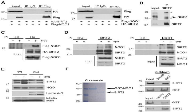
NQO1 interacts directly with SIRT2. (A) MCF-7 cells were transiently transfected with Flag-NQO1 and HA-SIRT2. 48 h after transfection, cell lysates were subjected to immunoprecipitation (IP) using either a Flag (left) or an HA (right) antibody followed by western blotting using antibodies against HA and Flag, respectively. Specificity was confirmed by using species-matched control IgG as a negative control. (B) Cell lysates from MCF-7 cells were subjected to immunoprecipitation (IP) using a SIRT2 antibody followed by western blotting using an anti-NQO1 antibody. * denotes Ig heavy and light chain. (C) Cell lysates from MCF-7 cells as described in (A) either unsynchronized or synchronized in mitosis after treatment with nocodazole (Noc, 200 nM for 12 h) were subjected to immunoprecipitation using an antibody against HA followed by western blotting using a Flag antibody. Specificity was confirmed by using species-matched control IgG as a negative control. Overexpression of both Flag-NQO1 and HA-SIRT2 is confirmed by western blotting (lower). (D) Reciprocal co-immunoprecipitation of endogenous SIRT2 and NQO1 in either unsynchronized or synchronized in mitosis Hela cells. For cell synchronization, HeLa cells were serum starved for 48 h and then released into regular media for 16 h. Cell lysates were subjected to immunoprecipitation (IP) using either a SIRT2 (left) or an NQO1 (right) antibody followed by western blotting using antibodies against SIRT2 and NQO1. Specificity was confirmed by using species-matched control IgG as a negative control. (E) Either unsynchronized or synchronized in mitosis (syn, as described in C) MCF-7 cells were fractionated into cytoplasmic extracts (cyt) and nuclear extracts (nuc). Levels of both SIRT2 and NQO1 were detected by western blotting. Successful cellular fractionation was confirmed by western blotting using antibodies against markers such as Lamin A/C (nuc) and actin (cyt). (F) Recombinant human SIRT2 and GST-NQO1 were used to check protein-protein interaction in vitro. Coomassie staining shows the molecular weight of the two proteins. GST-NQO1 was immunoprecipitated followed by SIRT2 immunodetection of (upper). In addition, SIRT2 was immunoprecipitated first, followed by immunodetection of GST-NQO1 (lower). Beads alone were used in the pull-down assays as negative controls.
![NQO1 positively regulates SIRT2 activity. (A) MCF-7 cells were transfected with either control (ctr) siRNA or NQO1 siRNA. 48 h after transfection, lysates were analyzed by western blotting using antibodies against Lys-40 acetylated tubulin (Ac-Tubulin), NQO1, SIRT2 and tubulin/actin. Quantification of acetylated tubulin levels are presented, **p<0.01. (B) MDA-MB-231 cells (NQO1 null) were transfected with either empty vector (ctr), Flag-NQO1 or Flag-NQO1P187S. 48h after transfection, lysates were analyzed by western blotting using antibodies against Lys-40 acetylated tubulin (Ac-Tubulin), Flag, SIRT2 and tubulin/actin. Quantification of acetylated tubulin levels are presented, **p<0.01. (C) MCF-7 cells were treated with Dicoumarol (100 uM) for different times or with different concentrations as indicated for 24h. Lysates were analyzed by western blotting using antibodies against Lys-40 acetylated tubulin (Ac-Tubulin), SIRT2 and tubulin. Quantification of acetylated tubulin levels are presented, *p<0.05. (D, E, F) MCF-7 cells were transfected with either control (ctr) siRNA or NQO1 siRNA. Cells were lysed in extraction buffer, and NADtotal (NAD and NADH) (D) as well as NADH alone (E) were calculated. The ratio of NAD/NADH (F) is calculated based on the formula (NADtotal-NADH)/NADH. Data represent Mean ± SEM of three independent experiments, *P<0.05. (G) MCF-7 cells were transfected with either control (ctr) siRNA or NQO1 siRNA. 48 h after transfection, SIRT2 activity in these lysates was determined by using a fluorometric assay based on the deacetylation of a unique target peptide included in the kit (FLUOR DE LYS? SIRT2 Deacetylase Fluorometric Assay Kit). Data represent Mean ± SEM of three independent experiments, *P<0.05. (H) MCF-7 cells were transfected with either control (ctr) siRNA or NQO1 siRNA. NQO1 knockdown cells were next transfected with NQO1 cDNA or treated with NAD+ (0.5 mM). Cell extracts were analyzed by western blotting using antibodies against Acetylated-Tubulin (Ac-tubulin), NQO1, SIRT2 and actin/GAPDH. * denotes exogenously expressed Flag-NQO1, whereas ** denotes the expression of endogenous NQO1. (I) MCF-7 cells were transfected with either control (ctr) siRNA or SIRT2 siRNA in the presence or absence of exogenously expressed NQO1 or NQO1P187S. Cell extracts were analyzed by western blotting using antibodies against acetylated histone H4 lysine16 (Ac-H4K16), SIRT2, Flag (NQO1) and actin. Note that NQO1P187S cannot be detected as this mutation affects enzymatic activity by extremely decreasing stability due to ubiquitination and proteasomal degradation [44]. (J) Wild-type (Sirt2+/+) and Sirt2 knockout (Sirt2-/-) MEFs were transfected with either control (ctr) siRNA, Nqo1 siRNA or Flag-NQO1 cDNA. Cell extracts were analyzed by western blotting using antibodies against acetylated histone H4 lysine16 (Ac-H4K16), SIRT2, Flag (NQO1) and actin.](productimages/extendimages/pmc06170003__nihms-1506327-f0004.jpg)
NQO1 positively regulates SIRT2 activity. (A) MCF-7 cells were transfected with either control (ctr) siRNA or NQO1 siRNA. 48 h after transfection, lysates were analyzed by western blotting using antibodies against Lys-40 acetylated tubulin (Ac-Tubulin), NQO1, SIRT2 and tubulin/actin. Quantification of acetylated tubulin levels are presented, **p<0.01. (B) MDA-MB-231 cells (NQO1 null) were transfected with either empty vector (ctr), Flag-NQO1 or Flag-NQO1P187S. 48h after transfection, lysates were analyzed by western blotting using antibodies against Lys-40 acetylated tubulin (Ac-Tubulin), Flag, SIRT2 and tubulin/actin. Quantification of acetylated tubulin levels are presented, **p<0.01. (C) MCF-7 cells were treated with Dicoumarol (100 uM) for different times or with different concentrations as indicated for 24h. Lysates were analyzed by western blotting using antibodies against Lys-40 acetylated tubulin (Ac-Tubulin), SIRT2 and tubulin. Quantification of acetylated tubulin levels are presented, *p<0.05. (D, E, F) MCF-7 cells were transfected with either control (ctr) siRNA or NQO1 siRNA. Cells were lysed in extraction buffer, and NADtotal (NAD and NADH) (D) as well as NADH alone (E) were calculated. The ratio of NAD/NADH (F) is calculated based on the formula (NADtotal-NADH)/NADH. Data represent Mean ± SEM of three independent experiments, *P<0.05. (G) MCF-7 cells were transfected with either control (ctr) siRNA or NQO1 siRNA. 48 h after transfection, SIRT2 activity in these lysates was determined by using a fluorometric assay based on the deacetylation of a unique target peptide included in the kit (FLUOR DE LYS? SIRT2 Deacetylase Fluorometric Assay Kit). Data represent Mean ± SEM of three independent experiments, *P<0.05. (H) MCF-7 cells were transfected with either control (ctr) siRNA or NQO1 siRNA. NQO1 knockdown cells were next transfected with NQO1 cDNA or treated with NAD+ (0.5 mM). Cell extracts were analyzed by western blotting using antibodies against Acetylated-Tubulin (Ac-tubulin), NQO1, SIRT2 and actin/GAPDH. * denotes exogenously expressed Flag-NQO1, whereas ** denotes the expression of endogenous NQO1. (I) MCF-7 cells were transfected with either control (ctr) siRNA or SIRT2 siRNA in the presence or absence of exogenously expressed NQO1 or NQO1P187S. Cell extracts were analyzed by western blotting using antibodies against acetylated histone H4 lysine16 (Ac-H4K16), SIRT2, Flag (NQO1) and actin. Note that NQO1P187S cannot be detected as this mutation affects enzymatic activity by extremely decreasing stability due to ubiquitination and proteasomal degradation [44]. (J) Wild-type (Sirt2+/+) and Sirt2 knockout (Sirt2-/-) MEFs were transfected with either control (ctr) siRNA, Nqo1 siRNA or Flag-NQO1 cDNA. Cell extracts were analyzed by western blotting using antibodies against acetylated histone H4 lysine16 (Ac-H4K16), SIRT2, Flag (NQO1) and actin.


