


Recombinant Human SOX10 protein, MYC/DDK-tagged 
| Cat.No. : | SOX10-1565H |
| Product Overview : | Recombinant Human SOX10, fussed with MYC/DDK tag at C-terminal, was expressed in HEK293 cells. |
| Availability | July 29, 2026 |
| Unit | |
| Price | |
| Qty |
- Specification
- Gene Information
- Related Products
- Citation
- Download
| Species : | Human |
| Source : | HEK293 |
| Tag : | DDK&Myc |
| Description : | This gene encodes an integral membrane protein that is required for cytokine-induced regulation of the tight junction paracellular permeability barrier. Mutations in this gene are thought to be a cause of band-like calcification with simplified gyration and polymicrogyria (BLC-PMG), an autosomal recessive neurologic disorder that is also known as pseudo-TORCH syndrome. Alternative splicing results in multiple transcript variants. A related pseudogene is present 1.5 Mb downstream on the q arm of chromosome 5. |
| Form : | 25 mM Tris.HCl, pH 7.3, 100 mM glycine, 10% glycerol. |
| Molecular Mass : | 49.7 kDa |
| Purity : | > 80% as determined by SDS-PAGE and Coomassie blue staining |
| Notes : | Recombinant protein was captured through anti-DDK affinity column followed by conventional chromatography steps. |
| Concentration : | >50 ug/mL as determined by microplate BCA method |
| Publications : |
| Gene Name | SOX10 SRY (sex determining region Y)-box 10 [ Homo sapiens ] |
| Official Symbol | SOX10 |
| Synonyms | SOX10; SRY (sex determining region Y)-box 10; transcription factor SOX-10; DOM; dominant megacolon; mouse; human homolog of; WS2E; WS4; SRY-related HMG-box gene 10; dominant megacolon, mouse, human homolog of; PCWH; WS4C; MGC15649; |
| Gene ID | 6663 |
| mRNA Refseq | NM_006941 |
| Protein Refseq | NP_008872 |
| MIM | 602229 |
| UniProt ID | P56693 |
| Chromosome Location | 22q13.1 |
| Function | DNA binding; RNA polymerase II core promoter proximal region sequence-specific DNA binding; RNA polymerase II distal enhancer sequence-specific DNA binding; chromatin binding; identical protein binding; protein binding; transcription coactivator activity; transcription regulatory region DNA binding; |
| ◆ Recombinant Proteins | ||
| SOX10-15772M | Recombinant Mouse SOX10 Protein | +Inquiry |
| SOX10-6355C | Recombinant Chicken SOX10 | +Inquiry |
| SOX10-5675R | Recombinant Rat SOX10 Protein | +Inquiry |
| SOX10-517H | Recombinant Human SOX10 Protein, GST-tagged | +Inquiry |
| SOX10-8583M | Recombinant Mouse SOX10 Protein, His (Fc)-Avi-tagged | +Inquiry |
| ◆ Cell & Tissue Lysates | ||
| SOX10-1565HCL | Recombinant Human SOX10 293 Cell Lysate | +Inquiry |
Temporal activation of WNT/β-catenin signaling is sufficient to inhibit SOX10 expression and block melanoma growth
Journal: Oncogene PubMed ID: 32238882 Data: 2020/4/1
Authors: Rexhep Uka, Christian Britschgi, Olga Shakhova
Article Snippet:To identify novel SOX10-interacting proteins, a M010817 WCE was analyzed by nLc Orbitrap MS at CovalX (CovalX AG, Zürich, Switzerland).To identify novel SOX10-interacting proteins, a M010817 WCE was analyzed by nLc Orbitrap MS at CovalX (CovalX AG, Zürich, Switzerland).. M010817 were lysed in NE buffer (50 mM Tris pH 7.5, 150 mM KCl, 0.2 mM EDTA, 5 mM MgCl 2 , 20% Glycerol, 0.5 mM DTT) and then sent to CovalX for further analysis, along with purified SOX10 protein in a buffer of 25 mM Tris pH 7.3, 100 mM glycine, 10% glycerol, obtained from Creative BioMart (Creative BioMart Inc, Shirley, NY, USA).. Procedure at CovalX were as follows:Procedure at CovalX were as follows:
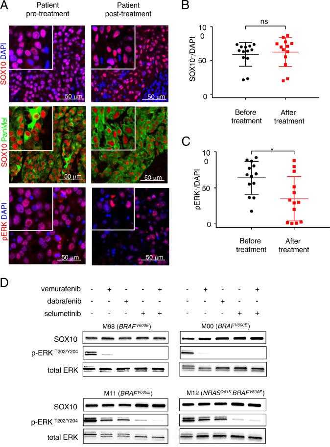
a Representative images of immunohistochemical stainings of human melanoma patient derived biopsies ( n = 13) before (left panel) and after (right panel) combined BRAF and MEK inhibitor treatment stained for
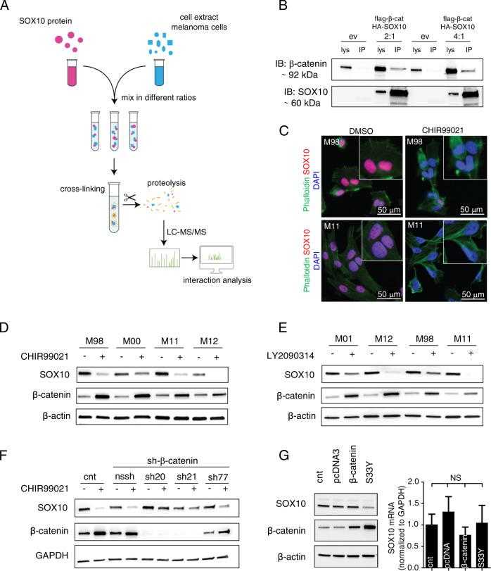
a Schematic representation showing the nLc Orbitrap mass spectrometry (MS) analysis to identify
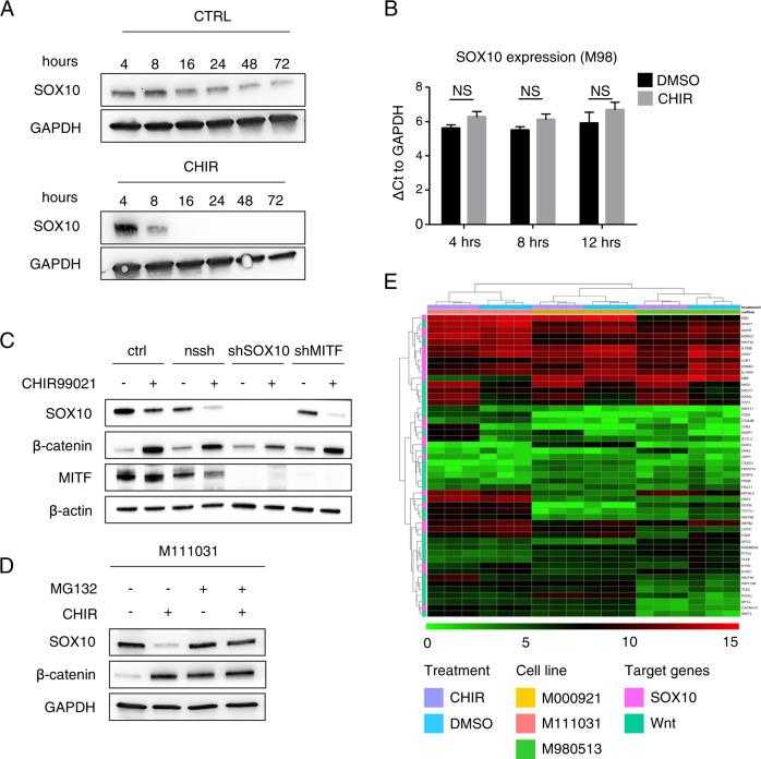
a Representative western blots of the indicated proteins in the MAPK inhibitor resistant human melanoma cell culture M12, at different timepoints of CHIR99021 (6 μM) treatment (timepoints are shown directly in the figure). GAPDH was used as loading control ( n = 3). b RNA levels in MAPK inhibitor sensitive (M98) human melanoma cell culture (ΔCt to GAPDH expression) after 4, 8, and 12 h of CHIR99021 (6 μM) treatment are shown compared with DMSO (ctrl) ( n = 3). c Representative western blot of the indicated proteins in the MAPK inhibitor sensitive human melanoma cell culture (M98). A representative western blot for the indicated proteins of M98 human melanoma cell culture stably expressing shSOX10 or shMITF constructs cultured in presence or absence of CHIR99021 for 24 h, 6 μM. Nssh represents the scrambled shRNA used as control construct ( n = 3). d Representative western blot of the indicated proteins in presence of CHIR99021 (6 μM) and/or MG-132 (20 μM) (in single or double treatments) for 16 h. GAPDH was used as a loading control ( n = 3). e Heatmap demonstrating the gene expression of
Not For Human Consumption!
Inquiry
- Reviews (0)
- Q&As (0)


Ask a Question for All SOX10 Products
Required fields are marked with *
My Review for All SOX10 Products
Required fields are marked with *