
SELE
-
Official Full Name
Selectin E -
Overview
E-selectin (Endothelial Leukocyte Adhesion Molecule-1, ELAM-1) belongs to the selectin family of adhesion molecules. Together with LECAM-1 (L-selectin) and GMP-140 (P-selectin), E-selectin mediates the initial interactions of leukocytes and platelets with endothelial cells. Molecular structure: The extracellular part of all selectins consists of an aminoterminal c-type lectin domain, which specifically binds to carbohydrate ligands. This is followed by an EGF-like domain, and, in the case of E-selectin, by 6 short consensus repeats. The transmembrane portion of the molecule is followed by a short cytoplasmic tail. Selectins provide the first, loose contacts of polymorphonuclear cells with the endothelium in areas of inflammation. The binding partner for E-selectin contains sialyl LewisX oligosaccharide or lactosaminoglycans that contain either sialic acid or fucose. E-selectin is expressed on cytokine-activated endothelial cells, and promotes the adhesion of leukocytes to the endothelium. This initial binding event is a prerequisite for the activation of the immune cells via inflammatory mediators. In contrast to GMP-140, E-selectin is maximally expressed 2-4 hours after cell activation. During the following 24-48 hours E-selectin is shed from the cytoplasmic membrane into the circulation. The circulating form of this selectin attracts neutrophils and activates the 2- integrins in preparing the cells for migration. Soluble E-selectin levels could provide insights into several pathologies, including allergic reactions, septic shock, vascular infection and inflammatory bowel disease. -
Synonyms
RP1-117P20.2;CD62E;ELAM;ELAM1;ESEL;LECAM2;CD62 antigen-like family member E;E-selectin;ELAM-1;endothelial adhesion molecule 1;endothelial leukocyte adhesion molecule 1;leukocyte endothelial cell adhesion molecule 2;leukocyte-endothelial cell adhesion molecule 2
Recombinant Proteins
- Human
- Rat
- Cynomolgus
- Mouse
- Dog
- Pig
- Rabbit
- Zebrafish
- Bovine
- HEK293
- Human Cells
- CHO
- C-His
- E.coli
- Mammalian Cells
- Insect Cells
- Yeast
- His
- Fc
- Non
- T7
- GST
- Flag
- Avi
- DDK
- Myc

Background
What is SELE protein?
SELE (selectin E) gene is a protein coding gene which situated on the long arm of chromosome 1 at locus 1q24. SELE is also known as CD62 antigen-like family member E (CD62E), endothelial-leukocyte adhesion molecule 1 (ELAM-1), or leukocyte-endothelial cell adhesion molecule 2 (LECAM2), a member of the Selectin family, is a 107-115 kDa cell surface glycoprotein. SELE consists of an N-terminal type 1 lectin domain, an EGF-like domain, 6 sushi (CCP/SCR) domains, a transmembrane sequence, and a short cytoplasmic domain.
What is the function of SELE protein?
During inflammation, SELE plays an important part in recruiting leukocytes to the site of injury. The local release of cytokines IL-1 and TNF-α by damaged cells induces the over-expression of SELE on endothelial cells of nearby blood vessels. SELE mediates the adhesion of tumor cells to endothelial cells, by binding to SELE ligands expressed by neutrophils, monocytes, eosinophils, memory-effector T-like lymphocytes, natural killer cells or cancer cells. Furthermore, a number of studies have reported that levels of SELE may be elevated in subjects with a variety of pathological conditions.
SELE related Signaling pathways
Multiple signaling pathways are involved in the regulation of SELE expression. One of the major pathways is the nuclear factor-kappa B (NF-κB) pathway, which is activated in response to various pro-inflammatory stimuli. Another important pathway involved in SELE regulation is the mitogen-activated protein kinase (MAPK) pathway. Furthermore, cytokines and chemokines released during inflammation, such as tumor necrosis factor-alpha (TNF-α) and interleukin-1 (IL-1), can also stimulate SELE expression through their respective signaling pathways.
SELE Related Diseases
Inflammatory bowel disease (IBD): SELE has been shown to be upregulated in patients with IBD, including Crohn's disease and ulcerative colitis. Cardiovascular diseases: SELE has been implicated in the development and progression of cardiovascular diseases, such as atherosclerosis, coronary artery disease, and ischemic stroke. Leukocyte adhesion deficiency Type I: This is a rare genetic disorder in which white blood cells cannot properly adhere to the lining of blood vessels due to a lack of the SELE protein. Sepsis: Sepsis is a life-threatening condition characterized by systemic inflammation in response to severe infection. Rheumatoid arthritis (RA): SELE is involved in the recruitment of leukocytes to the synovium, contributing to chronic inflammation in the joints of individuals with RA.
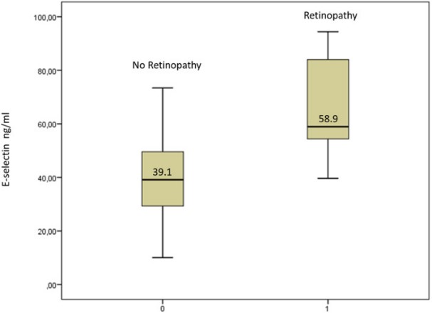
Fig1. E-selectin levels in sickle cell disease patients with and without retinopathy.
Bioapplications of SELE
SELE plays a critical role in various biological processes, including inflammation, immune response, cancer metastasis, and cardiovascular diseases. Understanding the signaling pathways involved in SELE expression and function can have important implications for therapeutic interventions and drug targeting strategies in these diseases.
Case Study
Case Study 1: Chae Min Lee, 2024
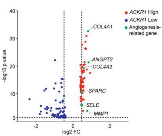
Fig1. Volcano plot illustrates several angiogenic process-related genes were enriched in ACKR1-high TECs.
Case Study 2: Sidra Shahid, 2022
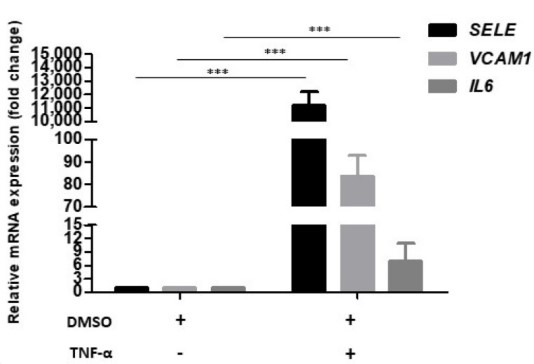
Fig2. Establishment of inflammatory phenotype in HUVECs. Bar graph showing relative mRNA levels of SELE, VCAM-I, and IL6 after normalization to housekeeping gene GAPDH, in DMSO control and TNF-α treated cells.
Quality Guarantee
High Purity
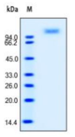
Fig1. SDS-PAGE (SELE-2242H) (PROTOCOL for western blot)
.
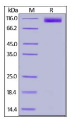
Fig2. SDS-PAGE (SELE-661H)
Involved Pathway
SELE involved in several pathways and played different roles in them. We selected most pathways SELE participated on our site, such as Cell adhesion molecules (CAMs),TNF signaling pathway,African trypanosomiasis, which may be useful for your reference. Also, other proteins which involved in the same pathway with SELE were listed below. Creative BioMart supplied nearly all the proteins listed, you can search them on our site.
| Pathway Name | Pathway Related Protein |
|---|---|
| African trypanosomiasis | GNAQ,IDO1,IL1B,IFNG,HPR,F2RL1,PLCB2,APOL1,IL10,IL6 |
| TNF signaling pathway | SOCS3,MMP3,PIK3CB,RPS6KA4,MAPK12,AKT2,MAP3K7,CXCL10,BAG4,CASP10 |
| Cell adhesion molecules (CAMs) | HLA-E,CLDN10A,CLDN7,HLA-DQB1,CDH1,MHC2DCB,LRRC4,BMA1,CLDN5A,CD6 |
| Malaria | TGFB1,TLR9,TGFB3,GYPC,KLRK1,SDC2,VCAM1,KLRC4-KLRK1,TLR4,SDC1 |
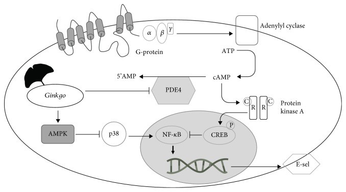
Fig2. Proposed mechanism to explain the effect of ginkgo extracts on signaling cascade involved in E-selectin regulation.
Protein Function
SELE has several biochemical functions, for example, oligosaccharide binding,phospholipase binding,protein binding. Some of the functions are cooperated with other proteins, some of the functions could acted by SELE itself. We selected most functions SELE had, and list some proteins which have the same functions with SELE. You can find most of the proteins on our site.
| Function | Related Protein |
|---|---|
| oligosaccharide binding | DCBLD1,CHID1,LOXL2,SELP,RS1A |
| phospholipase binding | CALM3,LMNB1,WAS,PRKCZ,CALM2,SNCA,APOC2,NEFL,PARK2,SERPINB1A |
| transmembrane signaling receptor activity | DNER,CLEC2D,OR4C12,LGR5,KLRD1,KLRC2,FCAMR,LILRA1,OR12D3,NFAM1 |
| protein binding | NCKIPSD,FIGNL1,ZBTB32,ZNF18,ZNF177,ADRB2,EVL,SPPL2B,ASMTL,SEC13 |
| sialic acid binding | AGRN,ST8SIA2,ADIPOQ,Siglece,SELP,FCN1,L1CAM,ST8SIA3 |
Interacting Protein
SELE has direct interactions with proteins and molecules. Those interactions were detected by several methods such as yeast two hybrid, co-IP, pull-down and so on. We selected proteins and molecules interacted with SELE here. Most of them are supplied by our site. Hope this information will be useful for your research of SELE.
PTPN11
Resources
Research Area
Related Services
Related Products
References
- Chukkapalli, SS; Rivera, MF; et al. Invasion of Oral and Aortic Tissues by Oral Spirochete Treponema denticola in ApoE(-/-) Mice Causally Links Periodontal Disease and Atherosclerosis. INFECTION AND IMMUNITY 82:1959-1967(2014).
- Przybyla, BD; Shafirstein, G; et al. Molecular changes in bone marrow, tumor and serum after conductive ablation of murine 4T1 breast carcinoma. INTERNATIONAL JOURNAL OF ONCOLOGY 44:600-608(2014).

