Recombinant Human NPC1 protein, His/FLAG-tagged
| Cat.No. : | NPC1-1339H |
| Product Overview : | Recombinant Human NPC1(Arg372-Phe622) fused with His/FLAG tag at N-terminal was expressed in HEK293. |
| Availability | January 29, 2026 |
| Unit | |
| Price | |
| Qty |
- Specification
- Gene Information
- Related Products
- Citation
- Download
| Species : | Human |
| Source : | HEK293 |
| Tag : | Flag&His |
| Protein Length : | Arg372-Phe622 |
| Predicted N Terminal : | His |
| Form : | Lyophilized from sterile PBS, pH 7.4.1. Normally 5 % - 8 % trehalose and mannitol are added as protectants before lyophilization. Specific concentrations are included in the hardcopy of COA.2. Please contact us for any concerns or special requirements. |
| Molecular Mass : | The recombinant human NPC1 consists 278 amino acids and predicts a molecular mass of 32 kDa. |
| Endotoxin : | < 1.0 EU per μg protein as determined by the LAL method. |
| Purity : | > 95 % as determined by SDS-PAGE |
| Stability : | Samples are stable for up to twelve months from date of receipt at -70 centigrade |
| Storage : | Store it under sterile conditions at -20 centigrade to -80 centigrade. It is recommended that the protein be aliquoted for optimal storage. Avoid repeated freeze-thaw cycles. |
| Reconstitution : | A hardcopy of COA with reconstitution instruction is sent along with the products. Please refer to it for detailed information. |
| Shipping : | In general, recombinant proteins are provided as lyophilized powder which are shipped at ambient temperature.Bulk packages of recombinant proteins are provided as frozen liquid. They are shipped out with blue ice unless customers require otherwise. |
| Publications : |
| Gene Name | NPC1 Niemann-Pick disease, type C1 [ Homo sapiens ] |
| Official Symbol | NPC1 |
| Synonyms | NPC1; Niemann-Pick disease, type C1; Niemann-Pick C1 protein; NPC; FLJ98532; |
| Gene ID | 4864 |
| mRNA Refseq | NM_000271 |
| Protein Refseq | NP_000262 |
| MIM | 607623 |
| UniProt ID | O15118 |
| Chromosome Location | 18q11-q12 |
| Pathway | Lysosome, organism-specific biosystem; Lysosome, conserved biosystem; |
| Function | hedgehog receptor activity; protein binding; receptor activity; sterol transporter activity; transmembrane signaling receptor activity; |
| ◆ Recombinant Proteins | ||
| NPC1-29133TH | Recombinant Human NPC1, GST-tagged | +Inquiry |
| NPC1-1339H | Recombinant Human NPC1 protein, His/FLAG-tagged | +Inquiry |
| NPC1-29132TH | Recombinant Human NPC1 | +Inquiry |
| NPC1-1338H | Recombinant Human NPC1 protein, His-tagged | +Inquiry |
| NPC1-4725H | Recombinant Human NPC1 Protein (Gln23-Asp266), N-His tagged | +Inquiry |
Multifunctional Pan-ebolavirus Antibody Recognizes a Site of Broad Vulnerability on the Ebolavirus Glycoprotein
Journal: Immunity PubMed ID: 30029854 Data: 2018/8/21
Authors: Pavlo Gilchuk, Natalia Kuzmina, James E. Crowe
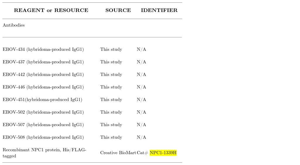
Article Snippet:Alexa Fluor 647 NHS ester , ThermoFisher , Cat# A37573.Alexa Fluor 647 NHS ester , ThermoFisher , Cat# A37573.. Recombinant NPC1 protein, His/FLAG-tagged , Creative BioMart , Cat# NPC1-1339H.. FluoSpheres NeutrAvidin-Labeled Microspheres , ThermoFisher , Cat# F-8776.FluoSpheres NeutrAvidin-Labeled Microspheres , ThermoFisher , Cat# F-8776.

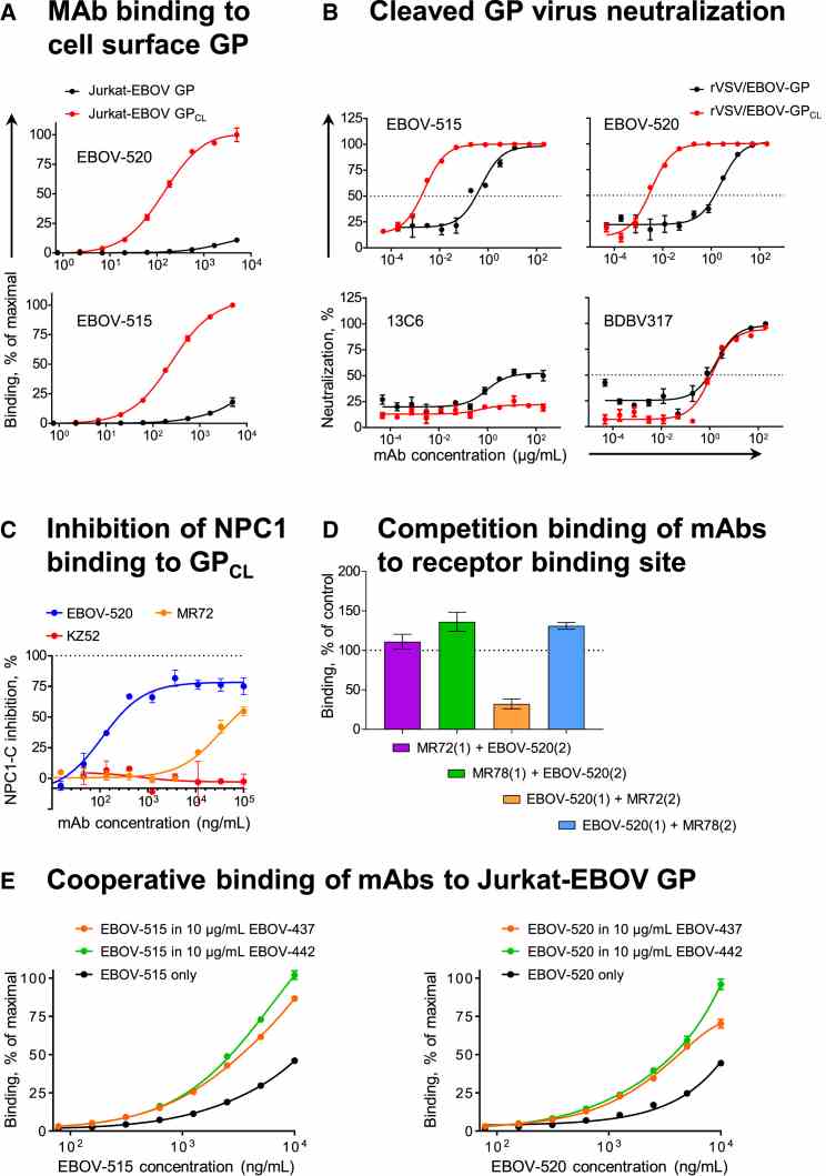
EBOV-515 and -520 Target Both Intact GP and Cleaved GP CL Intermediate to Neutralize the Virus (A) Binding curves for EBOV-515 or -520 using Jurkat-EBOV GP or Jurkat-EBOV GP CL . Fluorescently labeled mAbs were incubated with cells, and binding was assessed by flow cytometric analysis. (B) Neutralization curves for EBOV-515 or -520 or control mAbs 13C6 or BDBV317 using rVSV/EBOV-GP or rVSV/EBOV-GP CL . (C) Capacity of mAbs to inhibit

Not For Human Consumption!
Inquiry
- Reviews (0)
- Q&As (0)
Ask a Question for All NPC1 Products
Required fields are marked with *
My Review for All NPC1 Products
Required fields are marked with *


