Recombinant Rat CD38 Protein

Cat.No. : CD38-1249R
Product Overview : Recombinant Rat CD38 full length or partial length protein was expressed.
  • Specification
  • Gene Information
  • Related Products
  • Citation
  • Download
Species : Rat
Source : Mammalian Cells
Tag : His
Form : Liquid or lyophilized powder
Endotoxin : < 1.0 EU per μg of the protein as determined by the LAL method.
Purity : >80%
Notes : This item requires custom production and lead time is between 5-9 weeks. We can custom produce according to your specifications.
Storage : Store it at +4 ºC for short term. For long term storage, store it at -20 ºC~-80 ºC.
Storage Buffer : PBS buffer
Gene Name Cd38 CD38 molecule [ Rattus norvegicus ]
Official Symbol CD38
Gene ID 25668
mRNA Refseq NM_013127.1
Protein Refseq NP_037259.1
MIM
UniProt ID Q64244

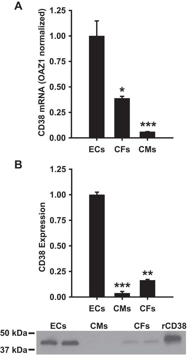
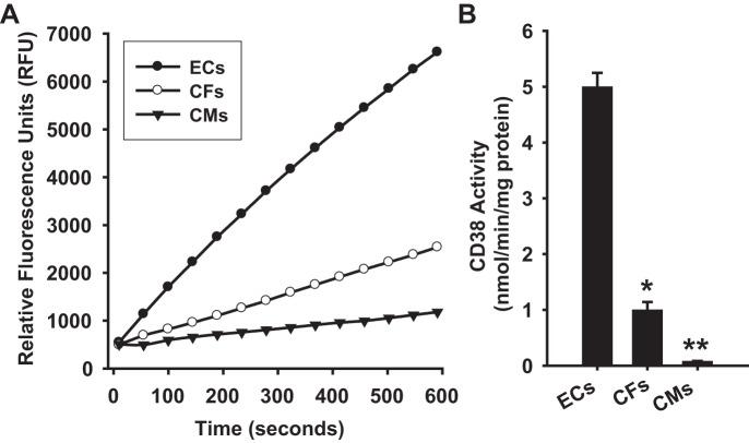
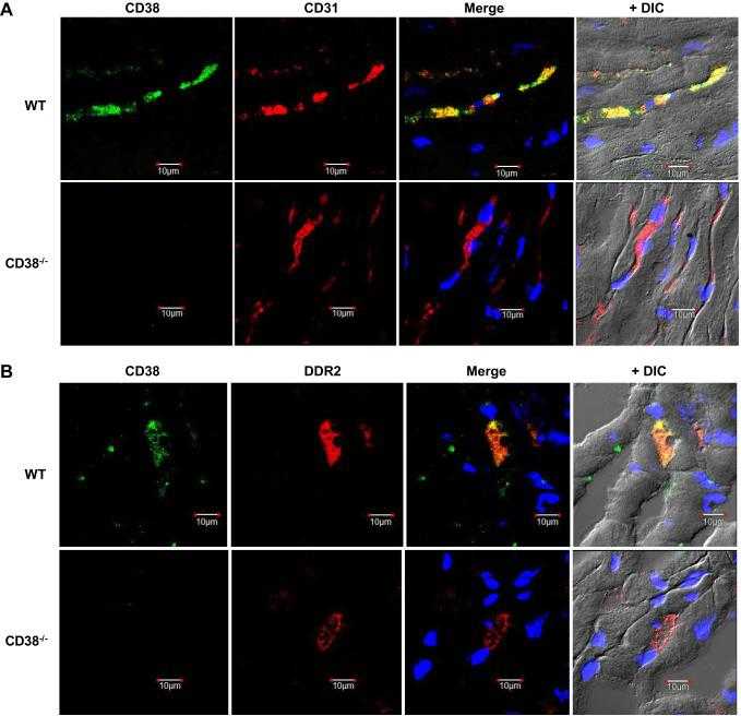
Characterization of CD38 in the major cell types of the heart: endothelial cells highly express CD38 with activation by hypoxia-reoxygenation triggering NAD(P)H depletion

Journal: American Journal of Physiology - Cell Physiology    PubMed ID: 29187364    Data: 2019/3/1

Authors: James Boslett, Craig Hemann, Jay L. Zweier

Article Snippet:ECL immunoblotting detection reagents were purchased from Amersham Biosciences.ECL immunoblotting detection reagents were purchased from Amersham Biosciences.. Rat recombinant CD38 was purchased from Creative BioMart.. Glass-bottomed 50-mm dishes for cell imaging were purchased from MatTek Corporation.Glass-bottomed 50-mm dishes for cell imaging were purchased from MatTek Corporation.

Cellular CD38 mRNA and expression levels. A: CD38 mRNA levels were significantly higher in ECs than in CFs and CMs. Levels were normalized to the housekeeping gene OAZ1, which showed similar levels of mRNA in each cell type. *P < 0.05 vs. ECs,***P < 0.001 vs. ECs (means ± SE; n = 4). B: Western blotting was used to determine the relative expression of CD38 in each cell type. For each cell type, 30 μg of total cellular protein was loaded to gels. The Western blot at the bottom of B shows a typical blot with 2 samples run for each cell type and on the right a standard of recombinant rat CD38 (rCD38) containing 100 ng of CD38. **P < 0.01 vs. ECs, ***P < 0.005 vs. ECs (means ± SE; n = 4).

Cellular CD38 mRNA and expression levels. A: CD38 mRNA levels were significantly higher in ECs than in CFs and CMs. Levels were normalized to the housekeeping gene OAZ1, which showed similar levels of mRNA in each cell type. *P < 0.05 vs. ECs,***P < 0.001 vs. ECs (means ± SE; n = 4). B: Western blotting was used to determine the relative expression of CD38 in each cell type. For each cell type, 30 μg of total cellular protein was loaded to gels. The Western blot at the bottom of B shows a typical blot with 2 samples run for each cell type and on the right a standard of recombinant rat CD38 (rCD38) containing 100 ng of CD38. **P < 0.01 vs. ECs, ***P < 0.005 vs. ECs (means ± SE; n = 4).

Cellular CD38 activity levels. A: CD38 NAD(P)+ase activity was measured in ECs, CFs, and CMs based on the increase in fluorescence with the conversion of NAD+ analog ε-NAD to the highly fluorescent product ε-ADPR. The signal is expressed as relative fluorescence units. From this signal the activity is calculated and expressed as nmol·min?1·mg protein?1. For each cell type, 100 μg of total cellular protein was used. B: in ECs, CD38 activity was found to be 5-fold higher than in CFs, and over 50-fold higher than in CMs. *P < 0.05 and **P < 0.01 vs. ECs (means ± SE; n = 4–6).

Cellular CD38 activity levels. A: CD38 NAD(P)+ase activity was measured in ECs, CFs, and CMs based on the increase in fluorescence with the conversion of NAD+ analog ε-NAD to the highly fluorescent product ε-ADPR. The signal is expressed as relative fluorescence units. From this signal the activity is calculated and expressed as nmol·min?1·mg protein?1. For each cell type, 100 μg of total cellular protein was used. B: in ECs, CD38 activity was found to be 5-fold higher than in CFs, and over 50-fold higher than in CMs. *P < 0.05 and **P < 0.01 vs. ECs (means ± SE; n = 4–6).

CD38, CD31, and DDR2 immunostaining in heart tissue. A: CD38 (green) and endothelial cell marker CD31 (red) were stained in WT and CD38?/? (KO) heart sections. Using a differential interference contrast (DIC) image, CD31 is seen to strongly and selectively stain vascular endothelium. CD38 colocalized with CD31 in the WT heart sections, with merging of the two images yielding a strong yellow color. As expected, no CD38 staining is seen in KO. B: CD38 (green) and fibroblast marker DDR2 (red) were stained in WT and CD38?/? heart sections (KO). Some colocalization of CD38 and DDR2 was observed in WT sections, with no CD38 in KO.

CD38, CD31, and DDR2 immunostaining in heart tissue. A: CD38 (green) and endothelial cell marker CD31 (red) were stained in WT and CD38?/? (KO) heart sections. Using a differential interference contrast (DIC) image, CD31 is seen to strongly and selectively stain vascular endothelium. CD38 colocalized with CD31 in the WT heart sections, with merging of the two images yielding a strong yellow color. As expected, no CD38 staining is seen in KO. B: CD38 (green) and fibroblast marker DDR2 (red) were stained in WT and CD38?/? heart sections (KO). Some colocalization of CD38 and DDR2 was observed in WT sections, with no CD38 in KO.

Not For Human Consumption!

Inquiry

  • Reviews (0)
  • Q&As (0)

Customer Reviews

Write a review

Ask a Question for All CD38 Products

Required fields are marked with *

My Review for All CD38 Products

Required fields are marked with *

0
cart-icon