


Protein L Agarose High Flow Resin
| Cat.No. : | COP25 |
- Specification
- Gene Information
- Related Products
- Download
| Tag : | Non |
| PRODUCT INFORMATION : | Product Overview: Protein L Agarose High Flow Resin belongs to the resins family for capture of antibodies and fragments containing kappa light chains, and is available with different pack sizes for easy purification, process development and scale up. Immunoprecipitation studies with Protein L Agarose High Flow are also commonly used to purify proteins or protein complexes. Description:Protein L Agarose High Flow Resin, with rProtein L coupled to high flow Agarose matrix, has been successfully used for a wide range of antibody and antibody fragments capture for its strong affinity to the variable region of antibody kappa light chains. Although it binds to the Fab portion of the immunoglobulin, Protein L does not interfere with the antigen-binding site of the antibody. Therefore, Protein L potentially can be used in immunoprecipitation (IP) procedures. As a good complementary choice for Protein A and Protein G, besides IgG, Protein L has been used for efficient purification of IgM, IgE, IgD, IgA and IgY. Protein L Agarose High Flow Resin also shows great advantages in purification of antibody fragments such as Fabs, single-chain variable fragments (scFv), and domain antibodies (Dabs). Features: Binding specificities that complement Protein A and G media. Suitable for different applications, including poly or monoclonal antibodies and antibody fragments from different species and subclasses for its broad binding specificity. Purification of monoclonal antibodies from FCS supplemented media without interference of bovine immunoglobulin. Single point attachment coupling chemistry gives better ligand accessibility for higher binding capacity. Specifications: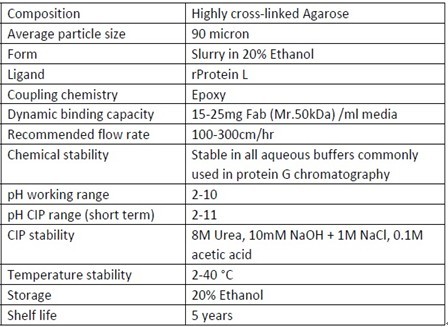 Note: Determined at 10% breakthrough at 4min residence time. Delivered at room temperature, and recommended long-term storage at 2-8°C. Note: Determined at 10% breakthrough at 4min residence time. Delivered at room temperature, and recommended long-term storage at 2-8°C. |
| INSTRUCTIONS FOR PURIFICATION : | 1. Column packing: Protein L Agarose High Flow is supplied as a suspension in 20% ethanol. Decant the 20% ethanol solution and replace it with water or other packing buffer required before use. Then follow the procedures below:(1). Equilibrate all material to the temperature at which the purification will be performed. Assemble the column (and packing device, if necessary).(2). Remove air from the column dead spaces by flushing the end-piece and adapter with packing buffer. Make sure no air has been trapped under the column net. Close the column outlet leaving the net covered with packing buffer.(3). Resuspend the medium stored in its container by shaking (avoid stirring the sedimented medium). Mix the packing buffer with the medium to form a 50% to 70% slurry (sedimented bed volume/total slurry volume = 0.5 to 0.7).(4). Pour the homogeneous slurry into the column in a single continuous motion. Pouring the slurry down a glass rod held against the column wall will help to minimize the introduction of air bubbles.(5). If using a packing device, immediately fill the remainder of the column and packing device with packing buffer. Mount the adapter or lid of the packing device and connect the column to a pump. Avoid trapping air bubbles under the adapter or in the inlet tubing.(6). Open the bottom outlet of the column and turn on the pump to run at the desired flow rate. Ideally, Protein L resin is packed at a constant pressure of approximately 1 bar (0.1 MPa). If the packing equipment does not include a pressure gauge, use a packing flow rate of approximately 400 cm/h (10 cm bed height, 25°C, water as packing buffer). If the recommended pressure or flow rate can not be obtained, use the maximum flow the pump can deliver.(7). When the bed height has stabilized, mark the compressed bed height and close the bottom outlet and stop the pump.(8). If using a packing device, disconnect the packing device and mount the adapter to the column.(9). With the adapter inlet open, push the adapter down, approximately 2 mm below previous compressed bed height, and the packing buffer will flush the adapter inlet. Close the adapter inlet.(10). The column is now ready to use for antibody purification.Note: Do not exceed 75% of the packing flow rate in subsequent chromatographic purification procedures.2. Purification2.1 Binding AffinityBinding characteristics of Protein L ligand are summarized in Table 2, which can be used as a general guide for affinity separation media selection of antibody purification.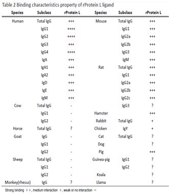 For purification of antibody fragments containing variable region of kappa light chain (VL-kappa), Protein L ligand also have be used successfully. Please refer to the following Figure 1 and Table 3 for separation media selection. For purification of antibody fragments containing variable region of kappa light chain (VL-kappa), Protein L ligand also have be used successfully. Please refer to the following Figure 1 and Table 3 for separation media selection.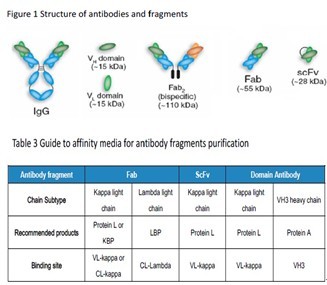 The dynamic binding capacity is a function of the sample residence time. It is necessary to use appropriate linear flow rate during sample application to ensure that residence time is in 4 to 8 min range at optimal column height of 5 to 20 cm.The residence time is equal to the packed bed height(cm) divided by the linear flow rate(cm/h) applied during sample loading.2.2 Purification parameters Generally, antibodies and fragments bind Protein L Agarose High Flow resin at neutral pH and physiological ionic strength, and are eluted at low pH. The recommended buffers for purification listed below can be used as good starting conditions for your experiments: (1). Binding buffer: 20mM Sodium phosphate, 150mM NaCl, pH7.2; 20mM Tris, 100mM NaCl, pH7-8; Phosphate buffered saline (PBS), pH 7.4 (0.01M phosphate buffer, 0.0027M KCl, 0.14M NaCl). (2). Elution buffer: 100mM Glycine, pH 2.0-3.5. (3). Neutralize buffer: 1M Tris pH 8-9. 2.3 Purification procedures1. Pack the column as described in "Column Packing" section. The recommended column height is within 5-20cm.2. Equilibrate the column at recommended flow rate with 5-10 column volumes of binding buffer to get a stable baseline.3. Calculate appropriate sample amount for loading. In principle, dynamic capacity is related to lots of parameters, such as antibody type, residence time, sample concentration, binding buffer and so on. Therefore, the maximum loading volume can be obtained by frontal analysis for individual sample under specific binding conditions. Generally, the dynamic binding capacity is higher than 15mg Fab /ml medium for 4-8min residence time.4. Apply clarified sample of antibody onto column. Samples need to be clarified by 0.45micron filter to remove any particles and colloids before application. It is recommended to dilute samples of high protein concentration, such as anti-serum, with equal volume of binding buffer to reduce sample viscosity.5. Wash column with 5 column volumes of binding buffer until UV level drop to baseline. Though not necessary for most of the cases, optional intermediate washing step with salts or detergents may help to remove impurities to some extent.6. Elute the column with 10 column volumes elution buffer. The most commonly used elution buffer is pH3.0; however, pH 2.5-3.0 is required for efficient elution of some kind of very strong binding antibodies with high recovery. Arginine and urea have been reported to improve antibody stability and avoid aggregation.7. Neutralize the elution peak immediately with 1M Tris buffer of pH 8.0-9.0.8. Re-equilibrate the column with 5-10 column volumes of neutral binding buffer. The dynamic binding capacity is a function of the sample residence time. It is necessary to use appropriate linear flow rate during sample application to ensure that residence time is in 4 to 8 min range at optimal column height of 5 to 20 cm.The residence time is equal to the packed bed height(cm) divided by the linear flow rate(cm/h) applied during sample loading.2.2 Purification parameters Generally, antibodies and fragments bind Protein L Agarose High Flow resin at neutral pH and physiological ionic strength, and are eluted at low pH. The recommended buffers for purification listed below can be used as good starting conditions for your experiments: (1). Binding buffer: 20mM Sodium phosphate, 150mM NaCl, pH7.2; 20mM Tris, 100mM NaCl, pH7-8; Phosphate buffered saline (PBS), pH 7.4 (0.01M phosphate buffer, 0.0027M KCl, 0.14M NaCl). (2). Elution buffer: 100mM Glycine, pH 2.0-3.5. (3). Neutralize buffer: 1M Tris pH 8-9. 2.3 Purification procedures1. Pack the column as described in "Column Packing" section. The recommended column height is within 5-20cm.2. Equilibrate the column at recommended flow rate with 5-10 column volumes of binding buffer to get a stable baseline.3. Calculate appropriate sample amount for loading. In principle, dynamic capacity is related to lots of parameters, such as antibody type, residence time, sample concentration, binding buffer and so on. Therefore, the maximum loading volume can be obtained by frontal analysis for individual sample under specific binding conditions. Generally, the dynamic binding capacity is higher than 15mg Fab /ml medium for 4-8min residence time.4. Apply clarified sample of antibody onto column. Samples need to be clarified by 0.45micron filter to remove any particles and colloids before application. It is recommended to dilute samples of high protein concentration, such as anti-serum, with equal volume of binding buffer to reduce sample viscosity.5. Wash column with 5 column volumes of binding buffer until UV level drop to baseline. Though not necessary for most of the cases, optional intermediate washing step with salts or detergents may help to remove impurities to some extent.6. Elute the column with 10 column volumes elution buffer. The most commonly used elution buffer is pH3.0; however, pH 2.5-3.0 is required for efficient elution of some kind of very strong binding antibodies with high recovery. Arginine and urea have been reported to improve antibody stability and avoid aggregation.7. Neutralize the elution peak immediately with 1M Tris buffer of pH 8.0-9.0.8. Re-equilibrate the column with 5-10 column volumes of neutral binding buffer. |
| PURIFICATION CASES : | Sample:Clarified Hybridoma cell culture containing mouse IgMBinding buffer: PBS pH 7.3Elution buffer: 100mM Glycine pH 3.0Column: Protein L Agarose High Flow Resin packed into a 0.5x 5 (cm, i.d. x Height) 1ml columnLoad sample volume: 100mlFlow rate: 1ml/min (300cm/hr) for loading and elutionUV280nm detection. Peak neutralized immediately with 1/20(v/v) 1M Tris pH8.0 after elution.Purity assay by 10-20% reduced gradient SDS-PAGE.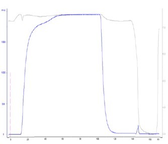 100 mL hybridoma cell culture was loaded onto Protein L Agarose High Flow column for mouse lgM capture. And finally lgM with >95% purity was obtained successfully within one step. 100 mL hybridoma cell culture was loaded onto Protein L Agarose High Flow column for mouse lgM capture. And finally lgM with >95% purity was obtained successfully within one step.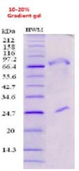 |
| CLEAN IN PLACE(CIP) : | Clean in place(CIP) is the important procedures for removing very tightly bound, precipitated or denatured proteins, DNA and Iipids, so as to maintain performance and capacity of the column.Protein L High Flow Resin allows the use of low concentration of NaOH as CIP agent, and recommended CIP procedures are as below: ClP procedures:(1). Wash the column with 3 to 5 column voIumes of binding buffer.(2). Backflush with 1 to 2 column voIumes of 8 M Urea or 10 mM NaOH with contact time of 10 minutes.(3). Wash immediately with 5-10 column volumes of binding buffer at pH 7-8 to remove ClP reagents.CIP is usually performed immediately after the elution. Before applying the alkaline NaOH CIP solution, we recommend equilibrating the column with a solution of neutral pH in order to avoid the direct contact between low pH elution buffer and high pH NaOH solution on the column. Mixing acid and alkaline solutions might cause a rise jn temperature in the column.Cleaning reagents concentration, contact time and frequency are typically the main parameters to vary during the optimization of the CIP. The nature of the feed material will ultimately determine the final CIP. However the general recommendation is to clean the column at least every 5 cycles during normal use. Depending on the nature of the contaminants, different protocols may have to be combined, for example 10 mM NaOH every cycle, and 8M Urea or 0.1 M glycine pH 2.5 every 10-20 cycles. 8M Urea can remove the precipitated proteins to restore the performance for resin. 1M NaCl can be introduced into CIP regents for stabilizing the ligand under alkaline conditions. |
| SANITIZATION : | Sanitization reduces microbial contamination of the chromatography column to a minimum. Protein L High Flow Resin allows the use of 0.1M acetate acid in 20% ethanol as sanitizing agent for sanitization.Sanitization procedures: 1. Wash the column with 3 column volumes of binding buffer.2. Wash with 0.1 M acetic acid in 20% ethanol for sanitization. Contact time of one hour is recommended.3. Wash immediately with at least 5 column volumes of sterile and filtered binding buffer at pH 7-8. |
| TROUBLE SHOOTING : | High column backpressure during purification1. Disconnect the column with system and make sure no tubings or connectors in the system caused the high system pressure; always use tubings and connectors of right inner diameters.2. Remove flow restrictor from systems if possible.3. Calibrate the pressure sensor in your systems.4. Make sure all buffers and samples be filtered through 0.22 or 0.45 micron disc membrane for clarification. For small volume sample, 10000g@10-20min centrifugation is an alternative solution.5. Lower flow rate when use buffers of high viscosity or working at cold temperature, especially during sample loading.6. Replace top screen net of column adapter in case of clogging.7. Lower the column bed height to 20 cm or less, too high beds will cause high pressure.8. Perform a thorough CIP procedure to restore the initial back pressure if column bed clogs. Unpack the column and wash media batch wise.9. Increase the CIP frequency and optimize the CIP regent formulations.10. Avoid freeze the medium or column during storage.Poor binding or low capacity1. Check the binding affinity of your antibodies of interest to the ligand.2. Make sure the pH values of binding buffer and sample are pH 7-8.3. Check if there exist some interference substances in binding buffer or samples, such as high concentration of chaotropic substances.4. Lower flow rate to give a residence time of 4-8min for sample loading.5. Check the history of the medium about how it has been cleaned and stored.Inefficient elution1. Check the pH value and composition for elution buffer.2. Try elution buffer of lower pH, for example pH 2.3.3. Use some chaotropic substances of low concentration in elution buffer.4. Introduce some solvents to decrease the polarity.5. Try other affinity media or other technologies.Low purity1. Reduce the sample holding time, lower purification temperature and always use protease inhibitors in samples and buffers to avoid degradation.2. Try to use as mild as possible elution conditions to avoid antibodies aggregation, and be sure to neutralize peak collected immediately after elution.3. Introduce an intermediate washing step before elution to remove any non-specific binding impurities, and some commonly used substances in washing buffer includes 1M NaCl, 0.5M Tetramethylammonium Chloride or detergents.4. Use pH linear 5-10 column volumes gradient (for example, phosphate and citrate to form pH 7.3 to 2.3 gradient) instead of stepwise elution and pool fractions of high purity.5. Alternative chromatography techniques need to be combined with affinity chromatography for higher purity with a multistep purification strategy, such as size exclusion and ionexchange, etc. |
| STORAGE : | Store unused media in its container at a temperature of 2 to 8°C for long term storage. Ensure that the container is closed and fully tightened. Equilibrate packed columns with 5-10 column volumes of 20% ethanol to prevent microbial growth. A thoroughly CIP procedure is recommended before long term storage. After storage, equilibrate with binding buffer and ready for use. Never freeze the media in case of generation of fine particles to increase the back pressure. |
| ◆ Recombinant Proteins | ||
| Ppl-728M | Recombinant Mouse Ppl Protein, His-tagged | +Inquiry |
| PPL-727H | Recombinant Human PPL Protein, His/GST-tagged | +Inquiry |
| PPL-2472H | Recombinant Human PPL protein(691-1260 aa), C-His-tagged | +Inquiry |
| PPL-3166H | Recombinant Human PPL Protein (Glu1439-Gly1716), His tagged | +Inquiry |
| PPL-7224Z | Recombinant Zebrafish PPL | +Inquiry |
Not For Human Consumption!
Inquiry
- Reviews (0)
- Q&As (0)


Ask a Question for All PpL Products
Required fields are marked with *
My Review for All PpL Products
Required fields are marked with *