A non-catalytic scaffolding activity of hexokinase 2 contributes to EMT and metastasis
Journal: Nature Communications PubMed ID: 35173161 Data: 2022/2/16
Authors: Catherine S. Blaha, Gopalakrishnan Ramakrishnan, Nissim Hay
Article Snippet:Enzyme-linked immunosorbent assays were conducted in Pierce Nickel-8-well-strip-coated plates.Enzyme-linked immunosorbent assays were conducted in Pierce Nickel-8-well-strip-coated plates.. The plates were coated with His-HK2 (50 nM; Creative BioMart) in coating buffer and incubated overnight at 4 °C.. Unbound proteins is then washed and blocking buffer is added for 1 h at room temperature.Unbound proteins is then washed and blocking buffer is added for 1 h at room temperature.
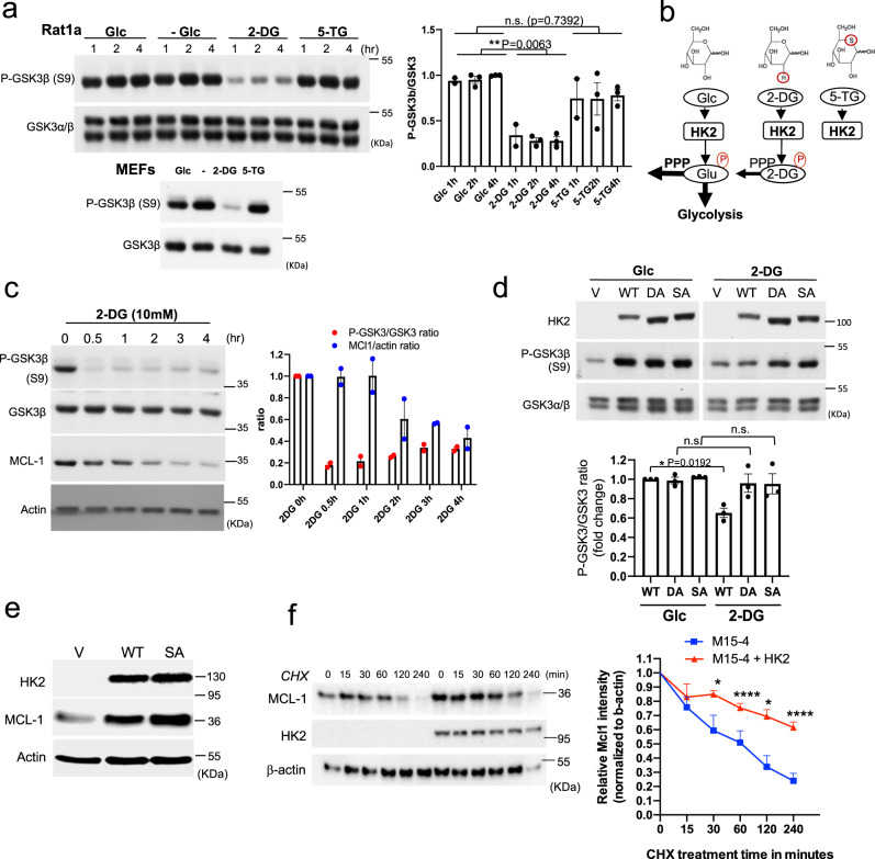
a Rat1a cells or MEFs were incubated in glucose-free medium in the absence (?) or presence of 10 mM glucose (Glc), 2-DG or 5-TG. Immunoblots showing GSK3β phosphorylation, at the indicated time points after incubation. Bar graph shows densitometric quantification of pGSK3β/GSK3α/β ratio from three independent experiments in Rat1a cells ( n = 3). Data are presented as the mean ± SEM. ** p = 0.0063; n.s. p = 0.7392; one-way ANOVA was used to calculate significance. b Schematic depicting the structures of glucose (Glc), 2-DG, and 5-TG and their utilization by HK inside cells. Like Glc, 2-DG can be phosphorylated by HK2 but cannot be further metabolized except in the first step of the pentose phosphate pathway (PPP). 5-TG cannot be phosphorylated by HK2. c Rat1a cells were incubated in glucose-free medium in the presence of 10 mM 2-DG for the indicated durations. Cells were then harvested for immunoblotting to determine GSK3β phosphorylation and MCL1 levels. Bar graph shows quantification from two independent experiments. d MI5-4 CHO cells expressing either wild-type (WT) HK2, individual kinase-dead HK2 mutants (DA, SA) or empty vector (V) were incubated in glucose-free medium in the presence of 10 mM glucose (G) or 2-DG (D). After 2 h, cells were harvested and analyzed for immunoblotting. Bar graph shows relative densitometric quantification of pGSK3β/GSK3α/β ratio (WT, DA, SA). Results are the mean ± SEM of three independent experiments. * p = 0.0192; one-way ANOVA was used to calculate significance. e Representative immunoblot of three independent experiments showing the level of the MCL-1 protein after expression of WT or kinase-dead SA mutant HK2 in MI5-4 CHO cells. f Protein stability of MCL-1 in MI5-4 CHO cells and MI5-4 CHO cells expressing WT HK2 as measured after exposure to cycloheximide (CHX). Plot showing MCL1 protein half-life after quantification relative to β-actin. Data are presented as the mean ± SEM. * p < 0.05 ( p = 0.0272 for 30 min and p = 0.0151 for 120 min), **** p < 0.0001 versus 0 h; two-way RM ANOVA test was performed to calculate the significance ( n = 3 independent experiments). Uncropped blots are provided as a Supplementary Fig. . Source data are provided as a Source Data file.
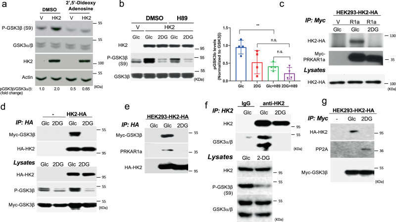
a Immunoblot showing GSK3β phosphorylation following HK2 expression in MI5-4 cells in the presence of the adenylate cyclase inhibitor 2’5’-dideoxyadenosine 200 μM for 6 h. Similar results obtained in two independent experiments. b GSK3β phosphorylation following HK2 expression in MI5-4 cells in the presence of the PKA inhibitor H89. Cells were pretreated with either DMSO or H89 (10 μM) for 2 h and then exposed to 10 mM glucose (Glc) or 2 DG for another 2 h in the presence of either DMSO or H89 (left panel: representative immunoblot; right panel: quantification of pGSK3β/GSK3β). Results are the mean ± SEM of three independent experiments. ** p = 0.008, n.s. = 0.1215; one-way ANOVA was used to calculate significance. c After transfection of control myc-tagged vector (V) or myc-tagged PRKAR1a (R1a) or control myc-vector plasmid into HEK293 cells stably expressing HA-tagged HK2 (HEK293-HK2-HA), cells were incubated in glucose-free medium in the presence of 10 mM glucose (Glc) or 2-DG. After 2 h, cells were lysed for immunoprecipitation with anti-myc antibody, followed by immunoblotting using anti-HA and anti-myc-HRP antibody. Total lysates immunoblotted using anti-HA antibody. Similar results obtained in two independent experiments. d Control or HEK293-HK2-HA cells were transfected with myc-GSK3β expression vector. Cells were treated, and lysed for immunoprecipitation as in c . Total lysates immunoblotted using anti-p-GSK3β, anti-myc-HRP, and anti-HA antibodies. Similar results obtained in two independent experiments. e After transfection of myc-GSK3β into HEK293-HK2-HA cells, cells were incubated in glucose-free medium in the presence of 10 mM glucose (Glc) or 2-DG. After 2 h, cells were lysed for immunoprecipitation with anti-HA antibody, followed by immunoblotting using anti-myc-HRP antibody and anti-PRKAR1a antibody. First lane shows control untransfected cells. Similar results obtained in two independent experiments. f HEK293 cells were incubated in glucose-free medium in the presence of 10 mM glucose (Glc) or 2-DG. After 2 h, cells were lysed for immunoprecipitation. Endogenous HK2 was immunoprecipitated with anti-HK2 and subjected to immunoblotting with anti-HK2 and anti-GSK3α/β. Similar results obtained in two experiments. g The experiment was performed as described in d , except that after immunoprecipitation with anti-myc, immunoblotting was performed with anti-PP2A. Similar results in two experiments. Uncropped blots are provided as a supplementary Fig. . Source data are provided as a Source Data file.
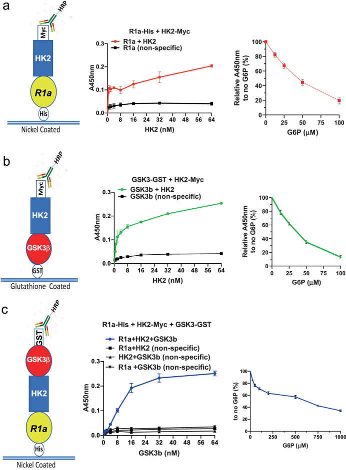
a Nickel-coated 96-well plates were incubated overnight at 4 °C with His-PRKAR1a (50 nM) and then incubated with Myc-HK2 (0.25–64 nM) in blocking buffer or with blocking buffer without HK2. HK2 binding was detected and quantified with anti-Myc-HRP-conjugated antibody, and an HRP-catalyzed reaction with a chromogenic substrate solution. Left panel shows generated binding curves. Curves were fitted based on a one-site-binding model in GraphPad Prism followed by a comparison of fits with p < 0.0001. Right panel: binding curve after increasing concentrations of G6P. Binding was conducted as above except that Myc-HK2 (32 nM) was incubated for 2 h. Thirty minutes before the end of Myc-HK2 incubation, increasing concentrations of G6P were added to the wells and detection was carried out as above. Results are the mean ± SEM of three independent experiments. p < 0.0001 one-way ANOVA. b Glutathione-coated 96-well plates were incubated overnight at 4 °C with GST-GSK3β (50 nM) and then incubated with or without Myc-HK2 (0.25–64 nM). Binding was quantified in a . Left panel: binding curves were generated as in a (comparison of fits with p < 0.0001). Right panel: binding curve after increasing concentrations of G6P as described in a . Results are the mean ± SEM of three independent experiments. p < 0.0001 one-way ANOVA. c Nickel-coated 96-well plates were incubated overnight at 4 °C with His-PRKAR1a (50 nM) or with blocking buffer in the absence of PRKAR1a, and then incubated with Myc-HK2 (32 nM) in blocking buffer or with blocking buffer without HK2 for 2 h. GST-GSK3β or blocking buffer without GSK3β was then added for 2 h. GSK3β binding was detected with anti-GST-HRP-conjugated antibody, and an HRP-catalyzed reaction with a chromogenic substrate solution. Left panel: binding curves were generated as in a (comparison of fits with p < 0.0001). Right panel: binding curve after increasing concentrations of G6P as described in a . Nickel-coated plates were successively incubated with His-PRKAR1a (50 nM) overnight, HK2-Myc (32 nM) for 2 h and then GST-GSK3β (32 nM) for 2 h. Right panel: results are the mean ± SEM of four independent experiments. panel one-way anova p < 0.0001.
A novel non-catalytic scaffolding activity of Hexokinase 2 contributes to EMT and metastasis
Journal: bioRxiv Data: 2021/5/10
Authors: Blaha Catherine, Ramakrishnan Gopalakrishnan, Hay Nissim
Article Snippet:PrePrint: Enzyme-linked immunosorbent assays were conducted in Pierce Nickel- 8-well-strip coated plates.Enzyme-linked immunosorbent assays were conducted in Pierce Nickel- 8-well-strip coated plates.. The plates were coated with His-HK2 (50nM; Creative BioMart) in coating buffer and incubated overnight at 4°C.. Unbound proteins is then washed and blocking buffer is added for 1h at room temperature.Unbound proteins is then washed and blocking buffer is added for 1h at room temperature.
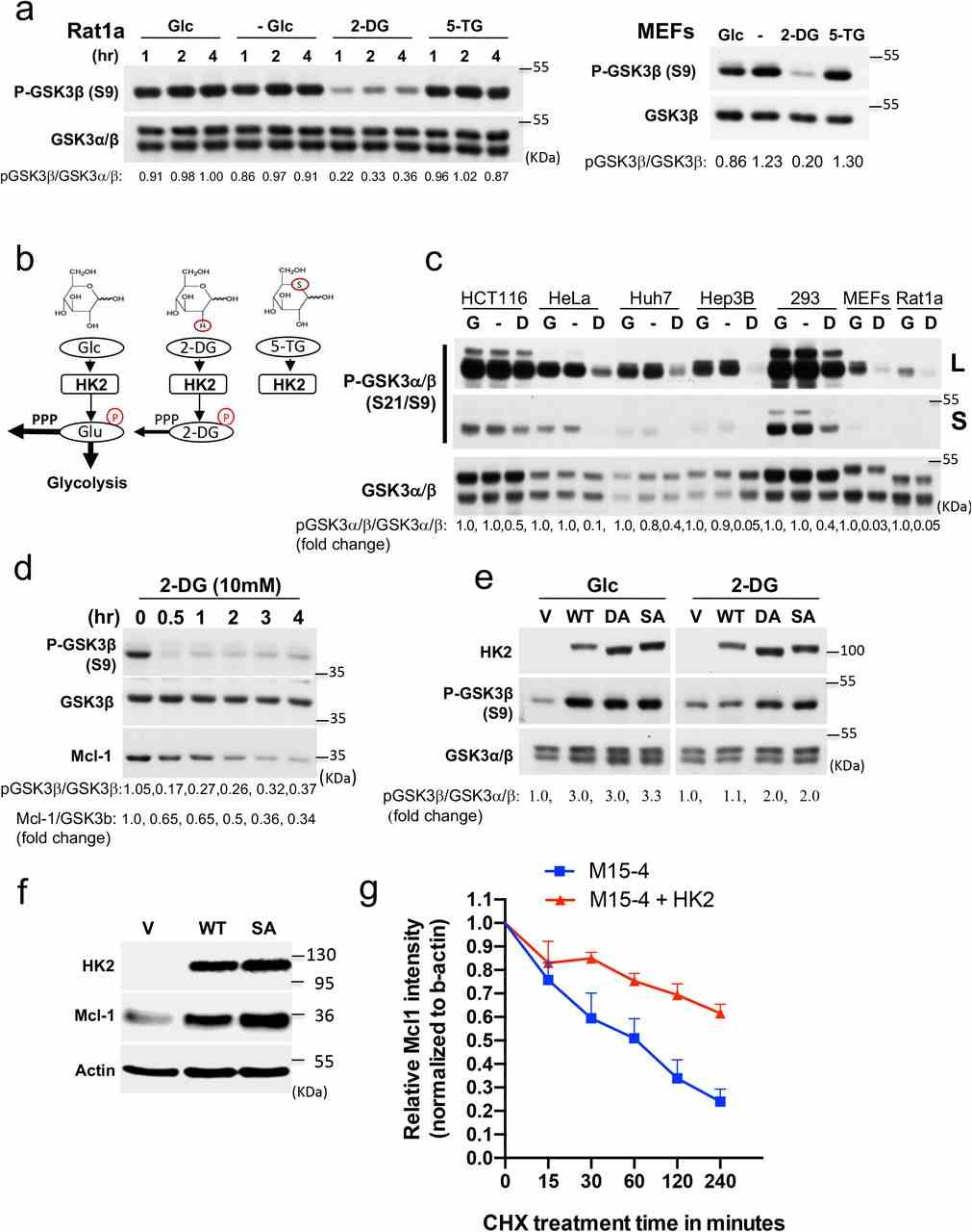
a. Rat1a cells or MEFs were incubated in glucose free medium in the absence (-) or presence of 10mM glucose (Glc), 2-DG or 5-TG. Immunoblots showing GSK3 β phosphorylation, at the indicated time points after incubation. b. Schematic depicting the structures of glucose (Glc), 2-DG, and 5-TG and their utilization by HK inside cells. Similar to Glc, 2-DG can be phosphorylated by HK2 but cannot be further metabolized except in the first step of the pentose phosphate pathway (PPP). 5-TG cannot be phosphorylated by HK2 c. Cells were incubated in the presence of glucose (G), absence of glucose (-) or presence of 2-DG (D) for 2 hr. An immunoblot image shows the phosphorylation of GSK3 α and GSK3 β . d. Rat1a cells were incubated in glucose-free medium in the presence of 10 mM 2-DG for the indicated durations. Cells were then harvested for immunoblotting to determine GSK3 β phosphorylation and MCL1 levels. e. MI5-4 CHO cells expressing either wild-type (WT) HK2, individual kinase-dead HK2 mutants (DA, SA) or empty vector (V) were incubated in glucose-free medium in the presence of 10 mM glucose (G) or 2-DG (D). After 2 hr, cells were harvested and analyzed for immunoblotting. f. The level of the Mcl-1 protein after expression of WT or kinase-dead SA mutant HK2 in M15- 4 CHO cells. g. Protein stability of MCL-1 in M15-4 CHO cells and M15-4 CHO cells expressing WT HK2 as measured after exposure to cycloheximide (CHX). Plot showing MCL1 protein half-life after quantification relative to b-actin in 3 independent experiments.
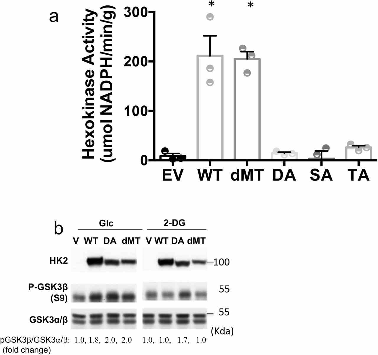
a. Hexokinase activity in M15-4 CHO cells expressing empty vector (EV), WT HK2 or HK2 mutants. Results are the mean ± SEM of 3 independent experiments in triplicate. *p<0.05, all 2-sided t- test vs. EV. b. MI5-4 CHO cells expressing either wild type (WT), kinase-dead HK2 mutant (DA), mitochondrial binding deficient mutant (dMT) or empty vector (V) were incubated in glucose free medium in the presence of 10mM glucose (G) or 2-DG (D). After 2hr, cells were harvested and analyzed by immunoblotting.
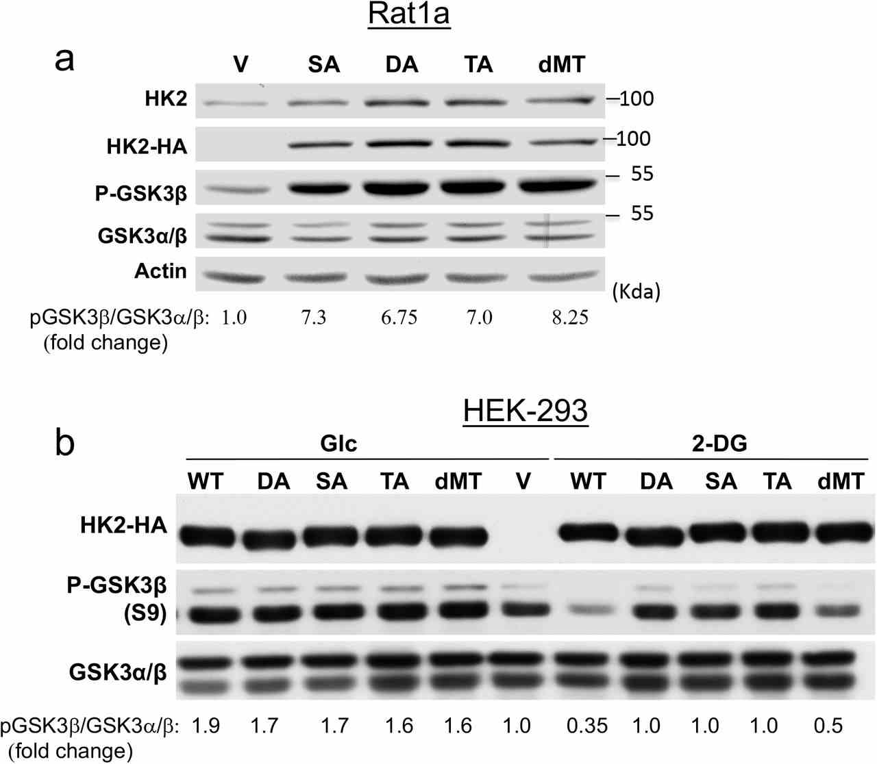
a. The effect of WT HK2 and HK2 mutants overexpression on GSK3β phosphorylation in Rat1a cells. b. The effect of 2-DG on GSK3β phosphorylation mediated by either WT HK2 or HK2 mutants in HEK293 cells.


