
Recombinant Human Klotho, Extracellular Domain, Fc Chimera
| Cat.No. : | KL-2155H |
| Product Overview : | Recombinant the signal peptide and the extracellular domain of human Klotho (aa 1-981) produced inHEK 293cells are fused at the C-terminus to the Fc portion of human IgG. |
- Specification
- Gene Information
- Related Products
- Citation
- Download
| Species : | Human |
| Source : | HEK293 |
| Tag : | Fc |
| Protein Length : | 1-981 a.a. |
| Description : | Klotho is a glycosylated protein that plays an important role in the regulation of phosphate and calcium homeostasis. Human Klotho exists in both membrane bound and secreted forms, and is predominantly expressed in the kidney convoluted tubules, and to a lesser extent, in the brain, reproductive organs, endocrine glands, urinary bladder, skeletal muscle, placenta, and colon. The full length transmembrane form has a large extracellular domain composed of two homologous subunits termed KL1 and KL2, which contain 516 and 439 amino acid residues, respectively, The predominant circulating form, which is derived from alternative RNA splicing, contains the KL1 subunit and constitutes the N-terminal sequence of transmembrane Klotho. A third Klotho protein of about 128 kDa has been identified in the blood and cerebrospinal fluid. |
| Purity : | ≥90% (SDS-PAGE). |
| Formulation : | Liquid. 0.2μm-filtered solution in PBS. |
| Concentration : | ~0.15mg/ml. |
| Endotoxin Content : | <1EU/µg protein (LAL-test). |
| Stability : | Working aliquots are stable for up to 3 months when stored at -20°C. |
| Gene Name | KL klotho [ Homo sapiens ] |
| Synonyms | KL; klotho; EC 3.2.1.31; Klotho peptide; OTTHUMP00000018796 |
| Gene ID | 9365 |
| mRNA Refseq | NM_004795 |
| Protein Refseq | NP_004786 |
| MIM | 604824 |
| UniProt ID | Q9UEF7 |
| Chromosome Location | 13q12 |
| Pathway | Signaling by FGFR |
| Function | beta-glucosidase activity; beta-glucuronidase activity; cation binding; fibroblast growth factor binding; hormone activity; protein binding; signal transducer activity; vitamin D binding |
CRIg Functions as a Macrophage Pattern Recognition Receptor to Directly Bind and Capture Blood-Borne Gram-Positive Bacteria.
Journal: Cell host & microbe PubMed ID: 27345697 Data: 2017/8/25
Authors: Zhutian Zeng, Bas G J Surewaard, Paul Kubes
Article Snippet:Bacteria trapped within Kupffer cells for more than five time points (2.5 min) were considered as captured.Bacteria trapped within Kupffer cells for more than five time points (2.5 min) were considered as captured.. Flow Cytometry of CRIg Binding to Bacteria Recombinant mouse or human extracellular domain of CRIg with a His-Tag (Creative BioMart) were labeled with AF 647 using a protein labeling kit according to the manufacturer’s instructions.. AF 647-labeled recombinant CRIg protein (20 mg/ml or indicated concentrations) was incubated with 2 3 105 CFU subcultured bacteria at room temperature for 2 to 3 hr with shaking.AF 647-labeled recombinant CRIg protein (20 mg/ml or indicated concentrations) was incubated with 2 3 105 CFU subcultured bacteria at room temperature for 2 to 3 hr with shaking.
Suppression of the Insulin Receptors in Adult Schistosoma japonicum Impacts on Parasite Growth and Development: Further Evidence of Vaccine Potential
Journal: PLoS Neglected Tropical Diseases PubMed ID: 25961574 Data: 2015/5/11
Authors: Hong You, Geoffrey N. Gobert, Mark Pearson
Article Snippet:Recombinant human insulin receptor (HIR) (short isoform) extracellular domain (Met1-Lys 956) (Creative BioMart, Inc., USA) and recombinant SjLD1 and SjLD2 (expressed in E . coli ) [ ] (see below) were subjected to electrophoresis on 12% (w/v) SDS-PAGE gels (1μg in 10μl SDS-PAGE sample buffer) as described above and then blotted onto a nitrocellulose membrane.(Met1-Lys 956) (Creative BioMart, Inc., USA) and recombinant SjLD1 and SjLD2 (expressed in E . coli ) [ ] (see below) were subjected to electrophoresis on 12% (w/v) SDS-PAGE gels (1μg in 10μl SDS-PAGE sample buffer) as described above and then blotted onto a nitrocellulose membrane. ... Rabbit anti-HIR polyclonal antibody (1:300) (Creative BioMart, Inc, Shirley, NY, USA), produced in a rabbit immunized with purified human cell-derived recombinant HIR extracellular domain (Met1-Lys 956), and the rabbit anti-SjLD1 (1:50) and anti-SjLD2 (1:50) sera were used as primary antibodies.. Then the membrane was incubated with anti-rabbit IgG-HRP (Invitrogen), diluted 1:3000, as described above.Then the membrane was incubated with anti-rabbit IgG-HRP (Invitrogen), diluted 1:3000, as described above.
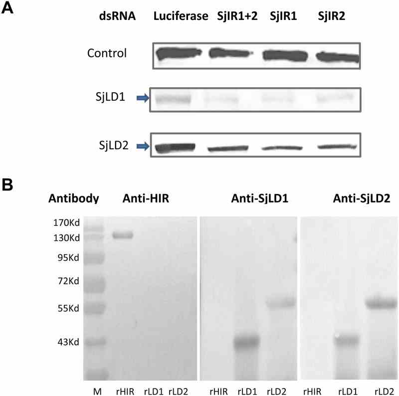
Results are shown for proteins recognised by anti-Sm-Pmy antibody (top panel), anti-SjLD1 (middle panel) and anti-SjLD2 (bottom panel) antibodies. The intensity of Sm-Pmy expression was evaluated so as to determine equal protein loading. The arrows indicate the diminished level of SjIR proteins relative to the luciferase treatment control in the first lane. The experiment was repeated twice with similar results obtained. B . Western blot analysis showing no immunological cross reactivity
Not For Human Consumption!
Inquiry
- Reviews (0)
- Q&As (0)


Ask a Question for All KL Products
Required fields are marked with *
My Review for All KL Products
Required fields are marked with *