


Active Recombinant Human TFPI protein, His-tagged 

| Cat.No. : | TFPI-875H |
| Product Overview : | Active Recombinant Human TFPI protein(NP_006278.1)(Asp29-Lys282) is expressed in HEK293, fused with a His tag at the C-terminus. |
| Availability | July 28, 2026 |
| Unit | |
| Price | |
| Qty |
- Specification
- Gene Information
- Related Products
- Citation
- Download
| Species : | Human |
| Source : | HEK293 |
| Tag : | His |
| Protein Length : | 29-282 aa |
| Form : | Lyophilized from a 0.22 μm filtered solution of PBS, pH 7.4. |
| Bio-activity : | Measured by its ability to inhibit trypsin cleavage of a fluorogenic peptide substrate, Mca-RPKPVE-Nval-WRK(Dnp)-NH2. The IC50 value is <0.41 nM. |
| Molecular Mass : | 30.01 kDa |
| Endotoxin : | < 0.1 EU/μg of the protein by LAL method. |
| Purity : | ≥ 95 % as determined by SDS-PAGE. |
| Storage : | Store at -20°C.Store the lyophilized protein at -20°C to -80°C up to 1 year from the date of receipt. After reconstitution, the protein solution is stable at -20°C for 3 months, at 2-8°C for up to 1 week. |
| Reconstitution : | Centrifuge the tube before opening. Reconstitute to a concentration of 0.1-0.5 mg/mL in sterile distilled water. Avoid vortex or vigorously pipetting the protein. For long term storage, it is recommended to add a carrier protein or stablizer (e.g. 0.1% BSA, 5% HSA, 10% FBS or 5% Trehalose), and aliquot the reconstituted protein solution to minimize free-thaw cycles. |
| Publications : |
| Gene Name | TFPI tissue factor pathway inhibitor (lipoprotein-associated coagulation inhibitor) [ Homo sapiens ] |
| Official Symbol | TFPI |
| Synonyms | TFPI; tissue factor pathway inhibitor (lipoprotein-associated coagulation inhibitor); LACI; tissue factor pathway inhibitor; EPI; extrinsic pathway inhibitor; TFI; TFPI1; anti-convertin; |
| Gene ID | 7035 |
| mRNA Refseq | NM_001032281 |
| Protein Refseq | NP_001027452 |
| MIM | 152310 |
| UniProt ID | P10646 |
| ◆ Recombinant Proteins | ||
| TFPI-646HB | Recombinant Human TFPI protein, His-Avi-tagged, Biotinylated | +Inquiry |
| TFPI-707H | Recombinant Human TFPI Protein, MYC/DDK-tagged | +Inquiry |
| TFPI-646H | Active Recombinant Human TFPI protein, His-Avi-tagged, Low Endotoxin | +Inquiry |
| Tfpi-575R | Active Recombinant Rat Tfpi protein, His-tagged | +Inquiry |
| TFPI-1246H | Recombinant Human TFPI Protein, His-tagged | +Inquiry |
| ◆ Cell & Tissue Lysates | ||
| TFPI-2746HCL | Recombinant Human TFPI cell lysate | +Inquiry |
| TFPI-2843MCL | Recombinant Mouse TFPI cell lysate | +Inquiry |
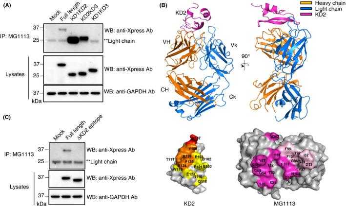
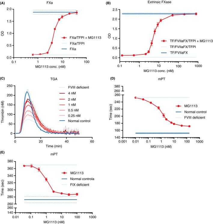
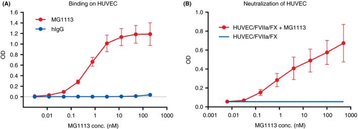
MG1113, a specific anti–tissue factor pathway inhibitor antibody, rebalances the coagulation system and promotes hemostasis in hemophilia
Journal: Research and Practice in Thrombosis and Haemostasis PubMed ID: 33313469 Data: 2020/10/22
Authors: Heechun Kwak, Sumin Lee, Sung Ho Hwang
Article Snippet:MG1113 and recombinant human (rh) TFPI (Creative BioMart, Shirley, NY, USA) were incubated at 37°C for 10 minutes. rhFXa (Enzyme Research Laboratories, South Bend, IN, USA) was then added and incubated at 37°C for 30 minutes, followed by the addition of FXa substrate, S‐2765 (Instrumentation Laboratory, Bedford, MA, USA).. The absorbance was measured using a VersaMax plate reader (Molecular Devices, San Jose, CA, USA) at 405 nm.The absorbance was measured using a VersaMax plate reader (Molecular Devices, San Jose, CA, USA) at 405 nm.
Neutralizing effect
Not For Human Consumption!
Inquiry
- Reviews (0)
- Q&As (0)


Ask a Question for All TFPI Products
Required fields are marked with *
My Review for All TFPI Products
Required fields are marked with *