

Active Recombinant Human TNFRSF17 protein, Fc-His-tagged 
| Cat.No. : | TNFRSF17-2348H |
| Product Overview : | Active Recombinant Human TNFRSF17 protein(NP_001183.2)(Met1-Ala54) is expressed in HEK293, fused with an Fc, His tag at the C-terminus. |
| Availability | October 06, 2026 |
| Unit | |
| Price | |
| Qty |
- Specification
- Gene Information
- Related Products
- Citation
- Download
| Species : | Human |
| Source : | HEK293 |
| Tag : | Fc&His |
| Protein Length : | 1-54 aa |
| Form : | Lyophilized from a 0.22 μm filtered solution of PBS, pH 7.4.Contact us for customized product form or formulation. |
| Bio-activity : | 1.Measured by its binding ability in a functional ELISA. Immobilized recombinant human BAFF at 5 μg/mL (100 μL/well) can bind recombinant human TNFRSF17 with a linear range of 3-20 ng/mL. 2.Loaded Human TNFRSF17/BCMA/CD269 Protein, C-hFc&His on ProA Biosensor, can bind Human TNFSF13B/BAFF/CD257 Protein, no Tag with an affinity constant of 2.62 nM as determined in BLI assay (Gator). |
| Molecular Mass : | 32.69 kDa |
| Endotoxin : | < 0.1 EU/μg of the protein by LAL method. |
| Purity : | ≥ 90 % as determined by SDS-PAGE. |
| Storage : | Store at -20°C.Store the lyophilized protein at -20°C to -80°C up to 1 year from the date of receipt. After reconstitution, the protein solution is stable at -20°C for 3 months, at 2-8°C for up to 1 week. |
| Reconstitution : | Centrifuge the tube before opening. Reconstitute to a concentration of 0.1-0.5 mg/mL in sterile distilled water. Avoid vortex or vigorously pipetting the protein. For long term storage, it is recommended to add a carrier protein or stablizer (e.g. 0.1% BSA, 5% HSA, 10% FBS or 5% Trehalose), and aliquot the reconstituted protein solution to minimize free-thaw cycles. |
| Gene Name | TNFRSF17 tumor necrosis factor receptor superfamily, member 17 [ Homo sapiens ] |
| Official Symbol | TNFRSF17 |
| Synonyms | TNFRSF17; tumor necrosis factor receptor superfamily, member 17; BCMA; tumor necrosis factor receptor superfamily member 17; BCM; CD269; B-cell maturation factor; B cell maturation antigen; B-cell maturation protein; |
| Gene ID | 608 |
| mRNA Refseq | NM_001192 |
| Protein Refseq | NP_001183 |
| MIM | 109545 |
| UniProt ID | Q02223 |
| ◆ Recombinant Proteins | ||
| TNFRSF17-027RP | Active Recombinant Rhesus TNFRSF17 protein, hFc-tagged, R-PE labeled | +Inquiry |
| TNFRSF17-7531H | Active Recombinant Human TNFRSF17 protein(R27P), His-tagged, Low Endotoxin | +Inquiry |
| Tnfrsf17-5074M | Recombinant Mouse Tnfrsf17 protein, hFc-tagged | +Inquiry |
| TNFRSF17-04D | Recombinant Dog BCMA Protein, His-tagged | +Inquiry |
| Tnfrsf17-1708M | Recombinant Mouse Tumor Necrosis Factor Receptor Superfamily, Member 17 | +Inquiry |

| ◆ Cell & Tissue Lysates | ||
| TNFRSF17-1489RCL | Recombinant Rat TNFRSF17 cell lysate | +Inquiry |
| TNFRSF17-2593MCL | Recombinant Mouse TNFRSF17 cell lysate | +Inquiry |
| TNFRSF17-1253CCL | Recombinant Cynomolgus TNFRSF17 cell lysate | +Inquiry |
| TNFRSF17-2489HCL | Recombinant Human TNFRSF17 cell lysate | +Inquiry |
Preclinical evaluation of CD8+ anti-BCMA mRNA CAR T-cells for treatment of multiple myeloma
Journal: Leukemia PubMed ID: 32632095 Data: 2020/6/26
Authors: Liang Lin, Shih-Feng Cho, Yu-Tzu Tai
Article Snippet:Viability of Descartes-08 cells was assessed using Acridine Orange and Propidium Iodide staining on the K2 Cellometer (Nexcelom Biosciences).Viability of Descartes-08 cells was assessed using Acridine Orange and Propidium Iodide staining on the K2 Cellometer (Nexcelom Biosciences).. CAR expression was determined by incubating CAR T-cells with 0.4 μg/ml of PE-conjugated recombinant BCMA (Recombinant human BCMA/TNFRSF17 protein, Fc/His-tagged, R-PE labeled; Creative BioMart, Shirley, NY) for 20 minutes at room temperature in FACS Buffer (DPBS, 0.5% BSA, 0.01% Sodium Azide).. Propidium Iodide was added at 1 μg/ml to stain non-viable cells.Propidium Iodide was added at 1 μg/ml to stain non-viable cells.
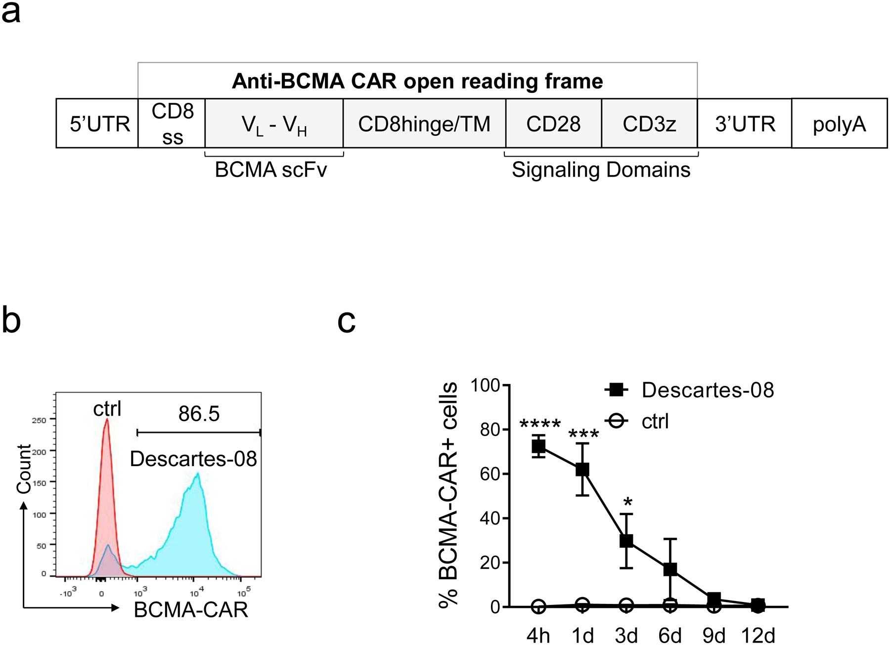
a Schematic representation of in vitro transcribed mRNA
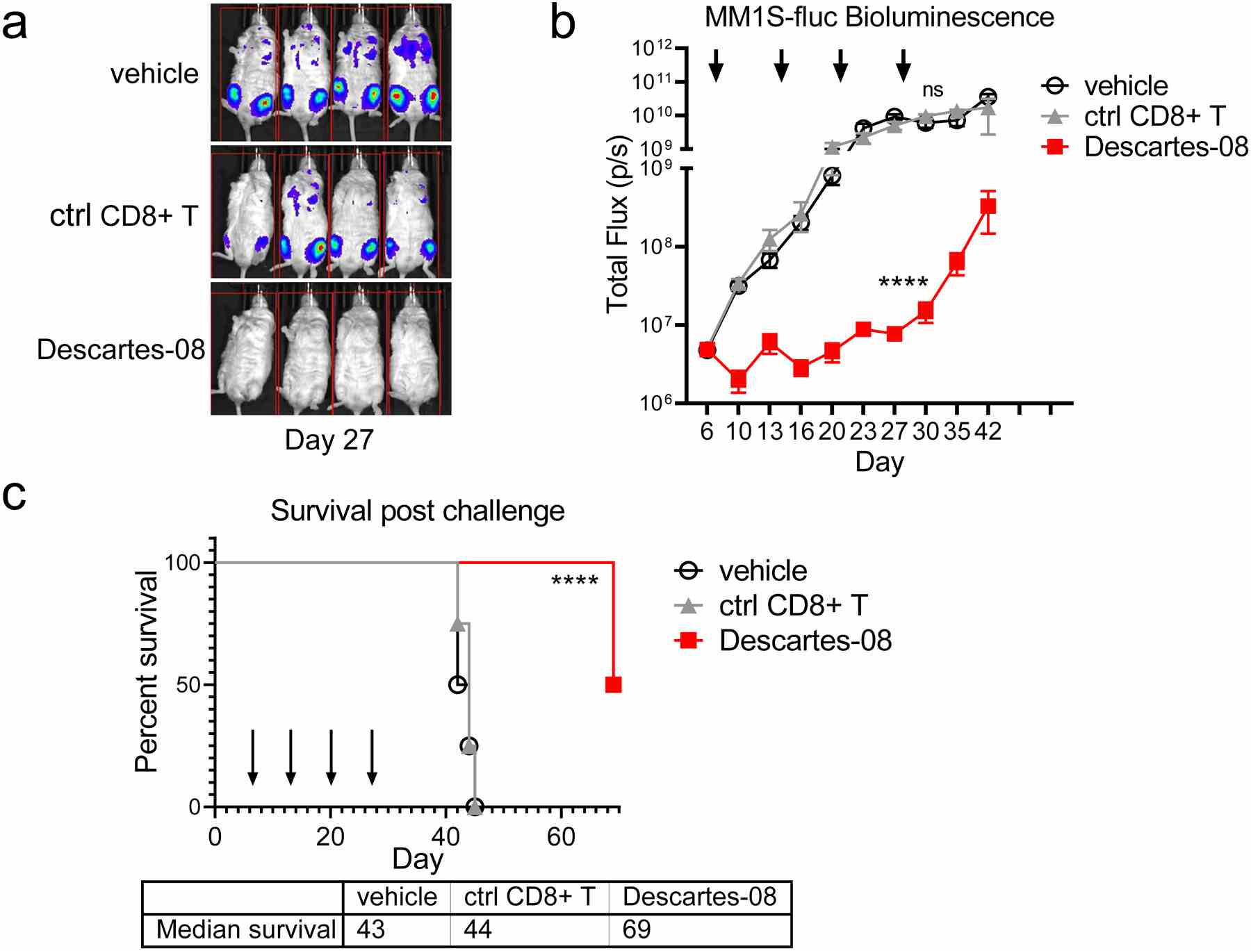
NSG mice receiving MM1S-luc cells (2 × 10 6 cells per mouse i.v.) at day 0 were randomized into treatment groups (n=4 per group) on day 6. On days 7, 14, 21 and 28, T-cells were thawed from cryovials, washed, and resuspended for administration via i.v. injection with vehicle, 20 × 10 6 mock-transfected control TCR knockout (KO) CD8+ (ctrl CD8+) or Descartes-08 cells (showing 67%

Not For Human Consumption!
Inquiry
- Reviews (0)
- Q&As (0)


Ask a Question for All TNFRSF17 Products
Required fields are marked with *
My Review for All TNFRSF17 Products
Required fields are marked with *
Recently Viewed