


Recombinant Human MANF protein 
| Cat.No. : | MANF-536H |
| Product Overview : | Recombinant Human MANF protein was expressed in Escherichia coli. |
| Availability | July 22, 2026 |
| Unit | |
| Price | |
| Qty |
- Specification
- Gene Information
- Related Products
- Citation
- Download
| Species : | Human |
| Source : | E.coli |
| Tag : | Non |
| Protein Length : | 158 |
| Description : | MANF is a secreted neurotrophic factor that is expressed in brain, neuronal and certain non-neuronal tissues. It has been shown to promote survival, growth and function of dopamine specific neurons. MANF and its structural homolog CDNF, each contain an N-terminal saposin-like lipid binding domain, and a carboxyl-terminal domain, which is not homologous to previously characterized protein structures. MANF and CDNF can prevent 6-OHDA induced degeneration of dopaminergic neurons by triggering survival pathways in a rat experimental model of Parkinson disease. Mature human MANF is 99 %, 98 % and 96 % a.a. identical to mature rat, mouse and bovine MANF respectively. |
| Form : | Lyophilized from a 0.2μm filtered concentrated solution in PBS, pH 7.4. |
| Bio-activity : | Fully biologically active when compared to standard. The ED50 as determined by a cell proliferation assay using rat C6 cells is less than 20 μg/ml, corresponding to a specific activity of > 50 IU/mg. |
| Molecular Mass : | Approximately 18.2 kDa, a single non-glycosylated polypeptide chain containing158 amino acids. |
| AA Sequence : | LRPGDCEVCISYLGRFYQDLKDRDVTFSPATIENELIKFCREARGKENRLCYYIGATDDAATKIINEVSKPLAHHIPVEKICEKLKKKDSQICELKYDKQIDLSTVDLKKLRVKELKKILDDWGETCKGCAEKSDYIRKINELMPKYAPKAASARTDL |
| Endotoxin : | Less than 1 EU/µg of rHuMANF as determined by LAL method. |
| Purity : | >95% by SDS-PAGE and HPLC analysis. |
| Storage : | Use a manual defrost freezer and avoid repeated freeze-thaw cycles. 12 months from date of receipt, -20 to -70 centigrade as supplied. 1 month, 2 to 8 centigrade under sterile conditions after reconstitution. 3 months, -20 to -70 centigrade under sterile conditions after reconstitution. |
| Reconstitution : | We recommend that this vial be briefly centrifuged prior to opening to bring the contents to the bottom. Reconstitute in sterile distilled water or aqueous buffer containing 0.1 % BSA to a concentration of 0.1-1.0 mg/mL. Stock solutions should be apportioned into working aliquots and stored at ≤-20 centigrade. Further dilutions should be made in appropriate buffered solutions. |
| Gene Name | MANF |
| Official Symbol | MANF |
| Synonyms | MANF; mesencephalic astrocyte-derived neurotrophic factor; arginine rich, mutated in early stage tumors , ARMET; ARP; arginine-rich, mutated in early stage tumors; ARMET; MGC142148; MGC142150; |
| Gene ID | 7873 |
| mRNA Refseq | NM_006010 |
| Protein Refseq | NP_006001 |
| MIM | 601916 |
| UniProt ID | P55145 |
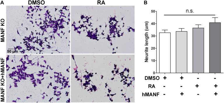
Mesencephalic Astrocyte-Derived Neurotrophic Factor (MANF) Regulates Neurite Outgrowth Through the Activation of Akt/mTOR and Erk/mTOR Signaling Pathways
Journal: Frontiers in Molecular Neuroscience PubMed ID: 33071755 Data: 2020/9/24
Authors: Wen Wen, Yongchao Wang, Jia Luo
Article Snippet:The following cells and materials were used: N2a (CCL-131), SH-SY5Y (CRL-2266) and HEK293 (CRL-1573) cells were from ATCC (Manassas, VA, United States); MEM (11095-080), high glucose DMEM (10569-010), L-methionine free DMEM (21013-024), FBS, antibiotic-antimycotic (15240112), GeneArt Genomic Cleavage Detection Kit (A24372), and Lipofectamine 3000 Reagent (L3000008) were from Life Technologies (Carlsbad, CA, United States); all- trans RA (R2625), crystal violet acetate (C5042), MTT (M5655), anhydrous DMSO (276855), DAPI (D9542), Akt activator SC79 (SML0749), and mTOR activator MHY1485 (SML0810) were from Sigma-Aldrich (St. Louis, MO, United States); PFA (15714) was from Electron Microscopy Sciences (Hatfield, PA, United States); recombinant human MANF (hMANF) (MANF-536H) was from Creative BioMart (New York, NY, United States); control CRISPR/Cas9 plasmid (sc-418922), mouse ARP double nickase plasmid (sc-428989-NIC), UltraCruz transfection reagent (sc-395739), plasmid transfection medium (sc-108062), and Akt inhibitor MK-2206 dihydrochloride (sc-364537) were from Santa Cruz Biotechnology (Dallas, TX, United States); Erk activator PDBu (12808), Erk inhibitor PD98059 (9900), and mTOR inhibitor Torin 1 (14379) were from Cell Signaling Technology (Beverly, MA, United States); pGEM-T-easy vector (A1360) was from Promega (Madison, WI, United States); 10-beta competent E. coli (C3019I) was from New England Biolabs (Ipswich, MA, United States); scrambled siRNA-GFP lentivector (LV015-G), Manf siRNA-GFP lentivector (279970940495), MANF-HA adenovirus (mouse) (279970540200), and CMV Null control adenovirus (000047A) were from Applied Biological Materials (Richmond, BC, Canada); PureLink Expi Endotoxin-free Maxi Plasmid Purification Kit (A31231) was from Thermo Fisher Scientific (Waltham, MA, United States); VECTASHIELD mounting medium (H-1400 and H-1500) was from Vector Laboratories (Burlingame, CA, United States); DC protein assay kit (5000112) was from Bio-Rad Laboratories (Hercules, CA, United States); Click-iT HPG Alexa Fluor Protein Synthesis Assay Kits (C10428) was from Invitrogen (Grand Island, NY, United States).. The following antibodies were used: anti-α-tubulin (T5168, Sigma-Aldrich); anti-ARMET/ARP (MANF) (ab67271 for C-terminus and ab67203 for N-terminus, Abcam, Cambridge, MA, United States); anti-HA-Tag (CST 3724), anti-phospho-Akt (Ser473) (CST9271), anti-Akt (CST9272), anti-phospho-Erk1/2 (CST 9101), anti-Erk1/2 antibody (CST 9102), anti-phospho-mTOR (Ser2448) (CST2971), anti-mTOR (CST2972), anti-phospho-p70 S6 (CST9204), anti-p70 S6 (CST2708), anti-Cas9 (CST 14697), and anti-β-Actin (CST3700) antibodies were all from Cell Signaling Technology; secondary antibodies conjugated to horseradish peroxidase (NA931V and NA934V) were from GE Healthcare Life Sciences (Pittsburgh, PA, United States); Alexa-488 conjugated anti-mouse (A21202), Alexa-594 conjugated anti-mouse (A11005) and Alexa-594 conjugated anti-rabbit antibodies (A11012) were from Life Technologies.
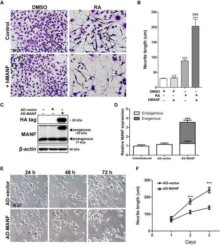
Addition of extracellular
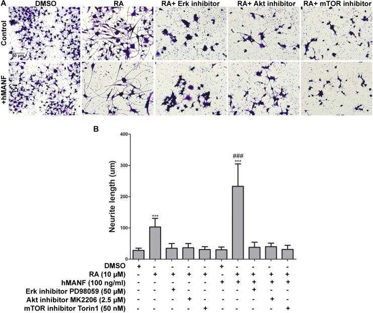
Pharmacological inhibition of Akt, Erk, and mTOR blocks
Not For Human Consumption!
Inquiry
- Reviews (0)
- Q&As (0)


Ask a Question for All MANF Products
Required fields are marked with *
My Review for All MANF Products
Required fields are marked with *