


Recombinant Mouse WT1 Protein
| Cat.No. : | WT1-18590M |
| Product Overview : | Recombinant Mouse WT1 full length or partial length protein was expressed. |
- Specification
- Gene Information
- Related Products
- Citation
- Download
| Species : | Mouse |
| Source : | Mammalian Cells |
| Tag : | His |
| Form : | Liquid or lyophilized powder |
| Endotoxin : | < 1.0 EU per μg of the protein as determined by the LAL method. |
| Purity : | >80% |
| Notes : | This item requires custom production and lead time is between 5-9 weeks. We can custom produce according to your specifications. |
| Storage : | Store it at +4 ºC for short term. For long term storage, store it at -20 ºC~-80 ºC. |
| Storage Buffer : | PBS buffer |
| Gene Name | Wt1 Wilms tumor 1 homolog [ Mus musculus ] |
| Official Symbol | WT1 |
| Gene ID | 22431 |
| mRNA Refseq | NM_144783.2 |
| Protein Refseq | NP_659032.3 |
| MIM | |
| UniProt ID | P22561 |
| ◆ Recombinant Proteins | ||
| WT1-589H | Recombinant Human WT1 Protein, His-tagged | +Inquiry |
| Wt1-1162M | Recombinant Mouse Wt1 protein, His&Myc-tagged | +Inquiry |
| WT1-07HFL | Recombinant Full Length Human WT1 Protein, N-His-tagged | +Inquiry |
| WT1-1402H | Recombinant Human WT1 protein, His & GST-tagged | +Inquiry |
| WT1-3769H | Recombinant Human WT1 protein, His-SUMO-tagged | +Inquiry |
| ◆ Cell & Tissue Lysates | ||
| WT1-278HCL | Recombinant Human WT1 293 Cell Lysate | +Inquiry |
WT1 Interacts with MAD2 and Regulates Mitotic Checkpoint Function
Journal: Nature communications PubMed ID: 25232865 Data: 2014/8/5
Authors: Jayasha Shandilya, Eneda Toska, Stefan GE Roberts
Article Snippet:GST, GST-WT1 (and deletion mutants) were expressed in BL21-DE3 competent cells, and His-tagged human MAD2 (and mutant derivatives) were expressed in BL21-DE3(pLysS) competent cells followed by affinity purification as described before .GST, GST-WT1 (and deletion mutants) were expressed in BL21-DE3 competent cells, and His-tagged human MAD2 (and mutant derivatives) were expressed in BL21-DE3(pLysS) competent cells followed by affinity purification as described before .. Flag-tagged full length WT1 protein (residue 1–449, including exon 5/17AA and KTS) was purchased from Creative BioMart.. The MAD2-binding peptide 1 (MBP1) was purchased from Peptide 2.0 (sequence: SWYSYPPPQRAV); Control peptide sequence: CKATKDPSRVGDS.The MAD2-binding peptide 1 (MBP1) was purchased from Peptide 2.0 (sequence: SWYSYPPPQRAV); Control peptide sequence: CKATKDPSRVGDS.
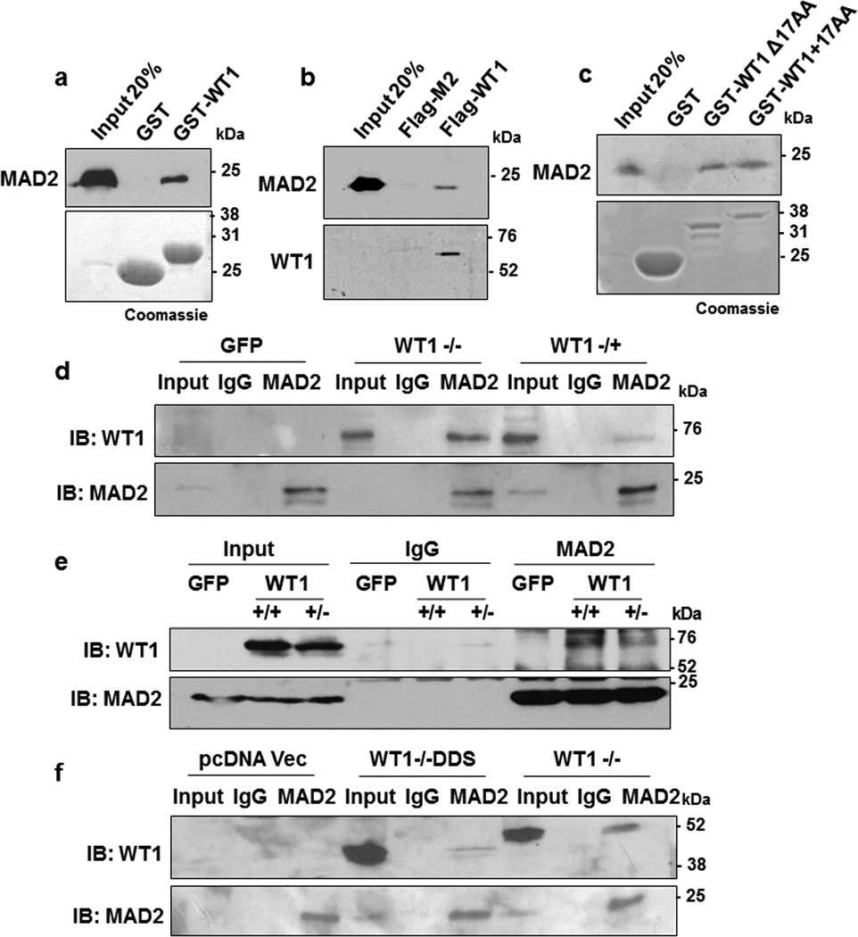
(a) In vitro interaction assay was performed with either
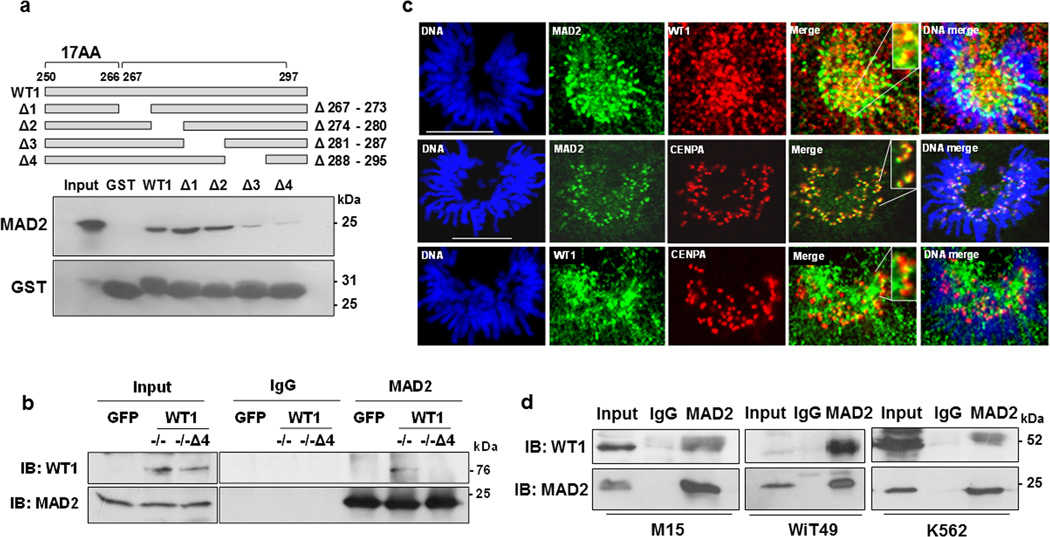
(a) GST-interaction assays were performed with the internal deletion mutants of
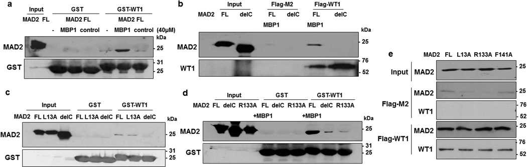
(a) GST-interaction assays were carried out with
Not For Human Consumption!
Inquiry
- Reviews (0)
- Q&As (0)


Ask a Question for All WT1 Products
Required fields are marked with *
My Review for All WT1 Products
Required fields are marked with *