

Active Recombinant Human CDH2 protein, His-tagged
| Cat.No. : | CDH2-2191H |
| Product Overview : | Active Recombinant Human CDH2 protein(NP_001783.2)(Met 1-Ala 724) was expressed in HEK293, fused with a polyhistidine tag at the C-terminus. |
| Availability | October 04, 2026 |
| Unit | |
| Price | |
| Qty |
- Specification
- Gene Information
- Related Products
- Case Study
- Application
- Download
| Species : | Human |
| Source : | HEK293 |
| Tag : | His |
| Protein Length : | 1-724 aa |
| Form : | Lyophilized from sterile PBS, pH 7.4. |
| Bio-activity : | Measured by the ability of the immobilized protein to support the adhesion of MCF-7 human breast adenocarcinoma cells. When 5 x 10E4 cells/well are added to Recombinant Human Cadherin-2 coated plates (5 μg/mL with 100 μL/well), apximately >50% will adhere after 1 hour at 37°C. |
| Molecular Mass : | The form of human CDH2 consists of 710 amino acids and predictes a molecular mass of 78.5 kDa. In SDS-PAGE under reducing conditions, the apparent molecular mass of CDH2 is 90 & 75 kDa corresponding to the fom and mature form respectinely due to glycosylation. |
| Endotoxin : | < 1.0 EU per μg of the protein as determined by the LAL method. |
| Purity : | > 95 % as determined by SDS-PAGE |
| Storage : | Store it under sterile conditions at -20°C to -80°C upon receiving. Recommend to aliquot the protein into smaller quantities for optimal storage. Avoid repeated freeze-thaw cycles. |
| Reconstitution : | It is recommended that sterile water be added to the vial to prepare a stock solution of 0.2 ug/ul. Centrifuge the vial at 4°C before opening to recover the entire contents. |
| protein Refseq-Weblink : | http://www.ncbi.nlm.nih.gov/protein/NP_001783.2 |
| Unit ID : | P19022 |
| Unit ID-Weblink : | http://www.uniprot.org/uniprot/P19022 |
| Gene Name | CDH2 cadherin 2, type 1, N-cadherin (neuronal) [ Homo sapiens ] |
| Official Symbol | CDH2 |
| Synonyms | CDH2; cadherin 2, type 1, N-cadherin (neuronal); NCAD; cadherin-2; CD325; CDHN; N cadherin; N-cadherin 1; neural cadherin; neural-cadherin; cadherin 2, N-cadherin (neuronal); calcium-dependent adhesion protein, neuronal; CDw325; |
| Gene ID | 1000 |
| mRNA Refseq | NM_001792 |
| protein Refseq | NP_001783 |
| MIM | 114020 |
| ◆ Recombinant Proteins | ||
| CDH2-44H | Recombinant Human CDH2 Protein, His (Fc)-Avi-tagged | +Inquiry |
| CDH2-87H | Recombinant Human Cadherin 2, Type 1, N-cadherin (Neuronal) | +Inquiry |
| RFL26809MF | Recombinant Full Length Mouse Cadherin-2(Cdh2) Protein, His-Tagged | +Inquiry |
| Cdh2-7025R | Recombinant Rat Cdh2 protein, His-tagged | +Inquiry |
| CDH2-3153H | Recombinant Human CDH2 Protein, His tagged | +Inquiry |

| ◆ Cell & Tissue Lysates | ||
| CDH2-971MCL | Recombinant Mouse CDH2 cell lysate | +Inquiry |
| CDH2-980HCL | Recombinant Human CDH2 cell lysate | +Inquiry |
Case 1: Sheehan SA, et al. Cell Commun Signal. 2022
Src kinase can stimulate tumor growth by activating podoplanin (PDPN), but nontransformed cells can counteract this through "contact normalization," requiring junctions with transformed cells. This study identified N-cadherin as crucial for this process. Experiments showed that cadherin-competent cells could restrict tumor growth, which cadherin-deficient cells couldn't do. RNA-seq indicated that cadherin's role in this mechanism affects around 10% of related gene transcripts, including PDPN. However, PDPN can push cells past this growth control, even when N-cadherin is present, showing its strong influence in bypassing contact normalization.
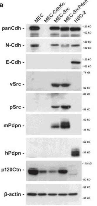
Fig1. Pan-Cdh, N-Cdh, E-Cdh, v-Src, active Src (phosphorylated at Tyr 416), mouse and human PDPN, p120-catnin (p120Ctn), and β-actin were detected by Western blot.
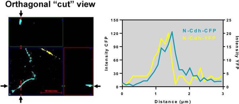
Fig2. Orthogonal imaging of N-Cdh-YFP and N-Cdh-CFP colocalization in cut out view.
Case 2: Choi S, et al. Theranostics. 2021
Bone marrow-derived mesenchymal stem cells (BM-MSCs) affect breast tumor behavior, partly through TGF-β signaling, but the exact role of movement-related molecules was unclear. This study showed that breast tumor cell media boosted BM-MSC migration by increasing N-cadherin expression, essential for maintaining cell adhesion. TGF-β enhances N-cadherin through Smad4, and disrupting N-cadherin or TGF-β pathways stopped BM-MSC movement towards tumor cells. This highlights N-cadherin’s key role in guiding BM-MSCs to breast tumors.
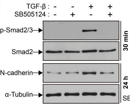
Fig1. Western blot analysis of phosphorylated Smad2/3 (p-Smad2/3), Smad2, N-cadherin.
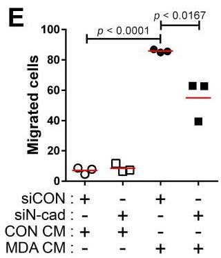
Fig2. Three-dimensional migration of BM-MSCs transfected with control siRNA (siCON) or N-cadherin siRNA (siN-cad).
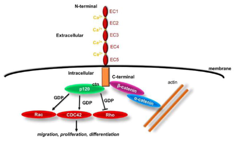
Fig1. Schematic representation of cadherin structure and downstream signaling. (Stella Alimperti, 2015)

Not For Human Consumption!
Inquiry
- Reviews (0)
- Q&As (0)


Ask a Question for All CDH2 Products
Required fields are marked with *
My Review for All CDH2 Products
Required fields are marked with *
Recently Viewed